
Salt Under a Microscope: What Salt Crystals Really Look Like
Sep 21st 2026
Salt may look like nothing more than tiny white grains when sprinkled onto food, but put those same grains under a microscope and they become surprisingly geometric.
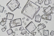
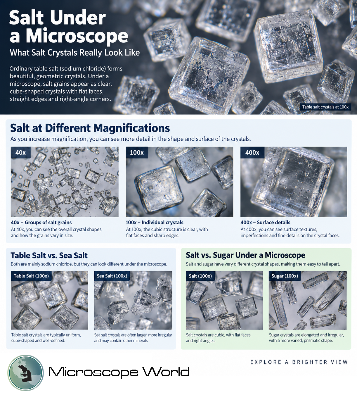
Under a microscope, table salt appears as transparent or translucent cube-shaped crystals with flat faces, straight edges and right-angle corners. Some grains form nearly perfect cubes, while others appear broken, stacked or irregular because of processing and handling.
Salt is also one of the easiest specimens to examine with a microscope. It does not require staining or complicated slide preparation, making it an excellent subject for students, classrooms and anyone learning how to use a microscope.
What Does Salt Look Like Under a Microscope?
When ordinary table salt is viewed under a microscope, its crystalline structure becomes much easier to see. Instead of looking like irregular white specks, individual grains reveal distinct geometric shapes.
Common features you may notice include:
- Square or cube-like shapes
- Straight, sharply defined edges
- Corners that meet at approximately 90-degree angles
- Flat crystal faces
- Transparent or translucent areas
- Broken edges and small fragments
- Crystals stacked on top of one another
Salt crystals are three-dimensional, but they may appear square rather than cubic when viewed directly from above. A traditional microscope provides a two-dimensional view of the specimen, so you may primarily see one face of the crystal.
Not every grain will look like a perfect cube, either. Grinding, packaging and handling can break crystals into smaller pieces. Scan around your sample and you will usually find several grains where the characteristic cubic structure is particularly easy to identify.
Why Are Salt Crystals Cube-Shaped?
The shape of salt isn't a coincidence. It comes from the structure of the material itself.
Ordinary table salt consists primarily of sodium chloride, or NaCl . Sodium chloride is made from positively charged sodium ions and negatively charged chloride ions.
In solid salt, these ions are arranged in an orderly, repeating three-dimensional crystal lattice. The sodium and chloride ions alternate throughout this structure.
This repeating arrangement produces sodium chloride's characteristic cubic crystal structure.
In other words, the large-scale geometry you observe under a microscope reflects the highly organized structure occurring at a scale far too small for an ordinary light microscope to resolve.
The American Chemical Society uses magnified salt crystals as an example for teaching this concept because the cubic shape can be observed in ordinary salt while models can demonstrate the arrangement of sodium and chloride ions within the crystal.
Salt Under a Microscope at Different Magnifications
One of the most interesting experiments is observing the same salt sample at progressively higher magnifications.
You do not need extreme magnification to see salt crystals. Their overall shapes can become apparent at relatively low power.
Salt at 40x Magnification
At 40x total magnification, you can view multiple grains simultaneously.
This is an excellent magnification for examining:
- Overall crystal shape
- Differences in grain size
- Groups of crystals
- Broken versus intact crystals
- Larger cubic structures
Starting at low magnification also makes it easier to locate and center an interesting grain before increasing the magnification.
Salt at 100x Magnification
At 100x, individual salt crystals become much more prominent.
You should be able to examine the flat faces, straight edges and right-angle corners that characterize sodium chloride crystals.
This is often an excellent magnification for observing salt because it provides a balance between detail and field of view. Microscope World has also photographed salt-containing crystal samples at 100x using polarized microscopy.
Salt at 400x Magnification
At 400x, you can concentrate on smaller portions of individual grains and examine finer surface features, fractures and imperfections.
However, higher magnification is not always better for viewing dry salt. Salt grains can be relatively thick compared with prepared biological specimens, so care should be taken when moving a high-power objective close to the slide.
For simply identifying the characteristic crystal shape of salt, lower magnifications are often sufficient.
How to View Salt Under a Microscope
Observing salt is a simple microscopy experiment that requires very little preparation.
What You'll Need
- Table salt
- Clean microscope slide
- Light microscope
- Small spoon, toothpick or tweezers
- Optional black paper for additional contrast when observing at very low magnification
Step 1: Prepare the Salt
Place a very small amount of dry table salt onto a clean microscope slide.
You only need a few grains. Using too much salt will create piles of overlapping crystals that can be difficult to examine.
Step 2: Start With Low Magnification
Place the slide onto the microscope stage and begin with your lowest-power objective.
Locate the salt grains and bring them into focus.
Starting at low power provides a larger field of view and makes finding individual crystals easier.
Step 3: Find a Well-Formed Crystal
Move the slide slowly while looking for an intact grain.
Look for crystals with:
- Four clearly visible sides
- Straight edges
- Right-angle corners
- Smooth or translucent faces
Center an interesting crystal within your field of view.
Step 4: Increase Magnification
Once the crystal is centered, increase the magnification.
Use the fine focus adjustment to sharpen the image.
As magnification increases, pay attention to the crystal's edges, surface texture, cracks and smaller crystals surrounding it.
Step 5: Adjust the Illumination
Salt can transmit light, so experimenting with the microscope's illumination and diaphragm may improve contrast.
Small lighting adjustments can make edges and surface features easier to distinguish.
Grow Salt Crystals and View Them Under a Microscope
You can take the experiment further by growing salt crystals yourself.
Instead of examining grains straight from the container, dissolve salt in water and allow the water to evaporate.
As the water disappears, sodium chloride comes out of solution and crystallizes again.
For a simple experiment:
- Add salt to warm water and stir until dissolved.
- Place a small drop of the saltwater onto a clean slide.
- Leave the slide undisturbed.
- Allow the water to evaporate completely.
- Examine the remaining crystals under low magnification.
You may find newly formed cubic crystals across the slide.
This experiment provides an interesting way to observe crystallization while demonstrating that the characteristic shape of salt is related to its underlying structure.
Table Salt vs. Sea Salt Under a Microscope
Table salt and sea salt are both composed primarily of sodium chloride, but they may not look identical under a microscope.
Table salt is generally processed to produce relatively consistent grains. Depending on the product, it may also contain iodine or anti-caking agents.
Under the microscope, you are likely to see many relatively uniform grains with recognizable cubic characteristics.
Sea salt is produced through the evaporation of seawater and may undergo less processing. Its grains can vary considerably in size and appearance and may also contain trace minerals.
As a result, a sea salt sample can contain larger and more irregular-looking grains than finely processed table salt.
Try placing small samples of table salt and sea salt on separate slides and viewing both at the same magnification. Comparing them side by side makes differences in crystal size and uniformity much easier to observe.
Salt vs. Sugar Under a Microscope
Salt and sugar may look similar in a kitchen, but their differences become much clearer under magnification.
Salt's cubic structure gives many grains a block-like appearance with straight edges and approximately 90-degree corners.
Sugar has a different crystal structure. Table sugar, or sucrose, tends to produce crystals that are more elongated, angled or irregular than sodium chloride.
|
Feature |
Salt |
Sugar |
|
Common compound |
Sodium chloride |
Sucrose |
|
Typical appearance |
Cubic or block-like |
Elongated or irregular |
|
Corners |
Often near 90 degrees |
More varied angles |
|
Crystal faces |
Flat, square-like |
More varied |
|
Appearance |
Transparent/translucent |
Transparent/translucent |
This makes salt vs. sugar under a microscope an easy comparison experiment for classrooms.
Place a few grains of each substance on separate slides and examine them using the same microscope and magnification.
Even though both appear as small white crystals without magnification, their different crystal structures become much more obvious when viewed closely.
Viewing Salt Crystals with Polarized Light
Salt and other crystals can also be investigated using specialized microscopy techniques.
Microscope World has previously examined mixtures containing sodium chloride using a polarizing microscope, including samples photographed at 40x and 100x magnification.
Polarized light microscopy is commonly useful when studying crystalline materials because interactions between polarized light and a specimen can reveal optical characteristics that may not be apparent with standard illumination.
If you're interested in taking crystal microscopy beyond this basic experiment, see our guide to Viewing Crystals with a Polarized Light Microscope .
Other Crystals to Examine Under a Microscope
Once you have examined salt, try comparing it with other crystalline substances.
Possible specimens include:
- Sugar
- Epsom salt
- Citric acid
- Baking soda
- Potassium chloride
The important thing to observe isn't simply whether a specimen contains crystals. Compare the shape, size, edges, angles, transparency and uniformity of the crystals.
These differences demonstrate how materials that appear similar to the naked eye can have dramatically different structures when magnified.
Frequently Asked Questions About Salt Under a Microscope
What does salt look like under a microscope?
Table salt typically appears as transparent or translucent cubic crystals. Individual grains can have flat faces, straight edges and approximately 90-degree corners. Broken grains may appear more irregular.
What shape is salt under a microscope?
Sodium chloride has a cubic crystal structure, so well-formed salt grains commonly appear cube-shaped or square when viewed from above.
Why is salt cube-shaped?
Salt's shape results from the repeating arrangement of sodium and chloride ions in its crystal lattice. This highly organized structure produces sodium chloride's characteristic cubic form.
What magnification do you need to see salt?
Salt can be observed at relatively low magnification. Starting around 40x allows you to view groups of crystals, while 100x provides a closer view of individual grains. Higher magnification can be used to investigate smaller surface details.
Can you see salt crystals at 40x?
Yes. Salt crystals are large enough that their overall shapes can be examined at 40x total magnification. Low magnification is particularly useful for comparing multiple grains and locating well-formed crystals.
What does salt look like at 100x?
At 100x magnification, individual salt grains become prominent, and their cubic characteristics are easier to identify. Look for flat crystal faces, straight edges and right-angle corners.
Can you view salt at 400x?
Yes, but 400x is not necessary for identifying salt's overall crystal shape. At higher magnification you can investigate smaller areas, fractures and surface imperfections. Care should be taken because dry salt grains can be thicker than conventional prepared specimens.
Does sea salt look different from table salt under a microscope?
It can. Both consist primarily of sodium chloride, but differences in processing, grain size and composition can affect their appearance. Sea salt may contain larger and less uniform grains than finely processed table salt.
How can you tell salt and sugar apart under a microscope?
Salt commonly forms cubic or block-like crystals with approximately right-angle corners. Sugar crystals generally have more elongated, angled or irregular shapes. Viewing the two samples side by side makes the difference particularly easy to see.
Can you see sodium and chloride ions with a light microscope?
No. A standard light microscope can reveal the larger salt crystal, but individual sodium and chloride ions are far too small to resolve. The cubic crystal you observe is made from an enormous number of ions arranged in a repeating structure.
Explore Crystals With a Microscope
Salt is an excellent example of how microscopy can reveal structures hidden in everyday objects. What appears to be an ordinary white grain to the naked eye becomes a remarkably organized geometric crystal under magnification.
And you don't need an advanced specimen or complicated preparation to see it. A few grains of table salt, a clean slide and a microscope are enough to begin exploring the microscopic structure of crystals.
From comparing salt and sugar to growing crystals through evaporation, these simple observations can be expanded into experiments for classrooms, laboratories and anyone interested in microscopy.


