
Diatoms Under a Microscope: What They Look Like and How to View Them
Nov 5th 2026
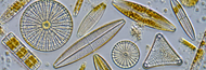
Diatoms are some of the most fascinating microorganisms to observe under a microscope. These microscopic algae have intricate, glass-like cell walls that form beautiful geometric patterns, making them popular subjects for microscopy enthusiasts, students, and researchers.
Whether you're examining pond water, studying aquatic ecosystems, or exploring microscopic structures, viewing diatoms under a microscope reveals details that are nearly impossible to appreciate with the naked eye.
What Are Diatoms?
Diatoms are single-celled algae found in freshwater, saltwater, and moist environments around the world. They play an important role in aquatic ecosystems by producing oxygen through photosynthesis and serving as a food source for other organisms.
What makes diatoms particularly interesting is their outer cell wall, called a frustule, which is primarily composed of silica, the same material found in glass.
These silica shells feature intricate patterns of pores, ridges, and other structures that vary among species. Scientists use these characteristics to identify and classify different types of diatoms.
Diatoms generally fall into two broad groups:
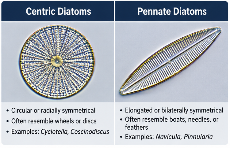
- Centric diatoms: Typically circular or radially symmetrical, often resembling tiny wheels or discs.
- Pennate diatoms: Usually elongated or bilaterally symmetrical, with shapes resembling boats, needles, or feathers.
Both types can be observed using a compound light microscope.
What Do Diatoms Look Like Under a Microscope?
Under a microscope, diatoms appear as tiny, transparent or golden-brown organisms with highly structured cell walls.
Their appearance depends on the species, magnification, and microscopy technique.
At lower magnifications, diatoms may resemble small oval, circular, or elongated shapes. As magnification increases, their distinctive silica shells become more visible.
Common features include:
- Geometric shapes: Circular, oval, triangular, and elongated forms.
- Fine patterns: Rows of tiny pores, lines, and ridges across the silica shell.
- Golden-brown coloration: Living diatoms often contain golden-brown chloroplasts.
- Transparent shells: Cleaned diatom frustules can appear nearly colorless under brightfield illumination.
Some pennate diatoms can also move slowly across surfaces through a process known as gliding motility.
The remarkable variety of diatom shapes and patterns makes them especially interesting for microscopic photography.
What Magnification Do You Need to See Diatoms?
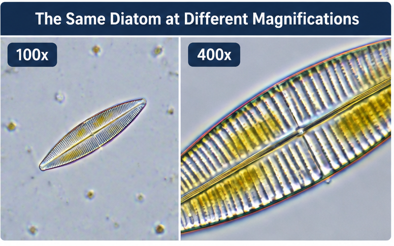
Most diatoms can be observed using a standard compound light microscope, although higher magnification is needed to examine their detailed structures.
|
Magnification |
What You Can See |
|
40x |
Larger diatoms and groups of diatoms, with limited detail |
|
100x |
General shapes and arrangements of many diatoms |
|
400x |
Individual diatoms, cell structures, and some shell patterns |
|
1000x |
Finer details of silica frustules, depending on optical resolution |
For most students and microscopy enthusiasts, 400x magnification provides an excellent balance between visibility and detail .
Researchers examining finer diatom structures may use 1000x magnification with an oil immersion objective. However, magnification alone does not guarantee better detail. Objective numerical aperture, illumination, and optical resolution are equally important.
Extremely fine structures may require scanning electron microscopy (SEM) to resolve.
How to View Diatoms Under a Microscope
Observing diatoms is relatively straightforward, especially when using prepared slides or samples collected from freshwater environments.
Step 1: Collect a Sample
Diatoms are commonly found in ponds, lakes, streams, and other aquatic environments.
Collect a small amount of water, preferably near submerged plants, rocks, or sediment where diatoms may be concentrated.
Alternatively, prepared diatom slides offer a convenient way to examine specimens without collecting samples.
Step 2: Prepare Your Slide
Place a small drop of your water sample onto a clean microscope slide.
Carefully lower a coverslip over the sample to minimize air bubbles.
Prepared slides containing cleaned diatom frustules are particularly useful for observing intricate silica structures.
Step 3: Start With Low Magnification
Begin with a 4x or 10x objective to locate diatoms within the sample.
Once you've identified an interesting specimen, center it in your field of view before increasing magnification.
Step 4: Increase Magnification
Switch to a 40x objective for a closer examination of the diatom's shape and internal structures.
Adjust the condenser and illumination to improve contrast.
Phase contrast or differential interference contrast (DIC) microscopy can help reveal transparent structures that may be difficult to distinguish using standard brightfield illumination.
Step 5: Photograph Your Diatoms
A digital microscope camera allows you to capture images of diatoms for identification, documentation, or comparison.
Photographing different specimens can also help demonstrate the remarkable diversity of diatom shapes and patterns.
What Is the Best Microscope for Viewing Diatoms?
A compound biological microscope is generally the best choice for observing diatoms because these microorganisms are small and often require magnifications between 100x and 1000x.
When choosing a microscope for diatom observation, consider the following features:
- Achromatic or plan objectives: Provide the optical quality needed to examine fine details.
- 400x to 1000x magnification: Suitable for viewing individual diatoms and many structural features.
- Adjustable LED illumination: Helps improve specimen visibility and contrast.
- Abbe condenser: Allows better control over illumination.
- Phase contrast capability: Useful for viewing transparent living specimens.
- Digital camera compatibility: Ideal for photographing and documenting diatom structures.
For educational and general observation, a quality compound microscope with 400x magnification is often sufficient. Advanced researchers may benefit from higher numerical aperture objectives and specialized contrast techniques.
Why Do Scientists Study Diatoms?
Diatoms are more than beautiful microscopic organisms. They provide valuable information about aquatic ecosystems and environmental conditions.
Scientists study diatoms for several reasons:
Water quality monitoring: Different diatom species respond to changes in nutrients, pollution, and environmental conditions, making them useful biological indicators.
Climate research: Preserved diatom shells in sediment layers help scientists investigate historical environmental and climate changes.
Ecological research: Diatoms are important primary producers in aquatic food webs and contribute significantly to global photosynthesis.
Microscopy research: Their intricate silica structures make diatoms useful specimens for evaluating microscope resolution and imaging techniques.
Frequently Asked Questions About Diatoms Under a Microscope
Can you see diatoms with a regular microscope?
Yes. Most diatoms can be viewed using a standard compound light microscope. Magnifications of 100x to 400x are suitable for observing their shapes, while higher magnifications reveal additional details.
Are diatoms visible to the naked eye?
Individual diatoms are generally too small to see without magnification. However, large accumulations of diatoms may appear as colored films or deposits on underwater surfaces.
Are diatoms alive when viewed under a microscope?
Diatoms collected from natural water samples may be alive. Living specimens often contain golden-brown chloroplasts, and some species exhibit slow gliding movements. Prepared slides may contain preserved specimens or cleaned silica shells.
What is the best magnification for diatoms?
For general observation, 400x magnification is an excellent starting point. Higher magnification, including 1000x, can help reveal finer structures when supported by suitable optics and illumination.
Explore the Microscopic World of Diatoms
Diatoms offer an incredible opportunity to explore the diversity and complexity of microscopic life. Their distinctive silica shells, geometric patterns, and ecological importance make them fascinating subjects for students, hobbyists, and professional researchers.
With a quality compound microscope and properly prepared samples, you can observe these remarkable microorganisms and discover details that are invisible to the naked eye.


