
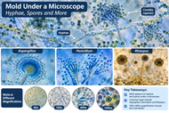
Mold Under a Microscope: What Mold Spores and Hyphae Look Like
Nov 12th 2026
Mold is a type of fungus that grows in damp environments, on food, and across many natural surfaces. While mold colonies may appear as fuzzy green, black, or white patches to the naked eye, looking at mold under a microscope reveals a much more complex structure.
Under magnification, mold can appear as networks of branching filaments, clusters of spores, and specialized structures that help fungi reproduce and spread.
Whether you're studying mold for a biology class, examining a prepared specimen, or working in a laboratory, microscopy provides a closer look at how these organisms grow.
What Does Mold Look Like Under a Microscope?
Under a microscope, mold typically appears as thin, threadlike structures called hyphae, along with round, oval, or other shaped spores. The appearance varies depending on the fungal species, its stage of development, and the magnification being used.
Common structures visible in mold specimens include:
- Hyphae: Long, branching filaments that form the main body of many molds.
- Mycelium: A network of interconnected hyphae that grows through or across a material.
- Spores: Microscopic reproductive structures that allow fungi to spread.
- Sporangia: Saclike structures that contain spores in certain molds.
- Conidiophores: Specialized stalks that produce or support asexual spores called conidia.
Some molds produce branching structures that resemble tiny trees, while others develop rounded spore-bearing structures at the ends of their filaments.
These features help scientists describe and differentiate fungi, although identifying a mold species generally requires more than visual inspection alone.
Mold Under a Microscope at Different Magnifications
The amount of detail visible depends on the microscope and magnification.
|
Magnification |
What You May See |
|
40x |
Larger fungal structures and the overall arrangement of a specimen |
|
100x |
Networks of hyphae, branching patterns, and some spore-bearing structures |
|
400x |
Individual spores, hyphal walls, and more detailed reproductive structures |
|
1000x |
Finer cellular details in suitably prepared specimens, typically using oil immersion |
For many educational observations, 100x to 400x total magnification provides a useful balance between field of view and structural detail.
At lower magnifications, it may be easier to examine the organization of fungal growth. Higher magnifications are useful for observing individual spores and distinguishing certain microscopic features.
Not every mold specimen requires 1000x magnification. The best setting depends on what you're trying to observe and how the sample has been prepared.
What Do Mold Spores Look Like Under a Microscope?
Mold spores are microscopic reproductive structures that vary considerably in shape, size, color, and arrangement.
Depending on the species, spores may appear:
- Round or spherical
- Oval or elongated
- Smooth or textured
- Transparent, pale, or darkly pigmented
- Arranged in chains, clusters, or enclosed structures
For example, some Aspergillus species produce chains of conidia around a specialized spore-bearing structure. Certain Penicillium species form branching, brushlike arrangements of conidia.
Other molds, including some Rhizopus species, develop rounded sporangia containing numerous spores.
Because different fungi can produce similar-looking structures, spore appearance alone is not sufficient to reliably identify a mold species or determine whether it is harmful.
Common Types of Mold Under a Microscope
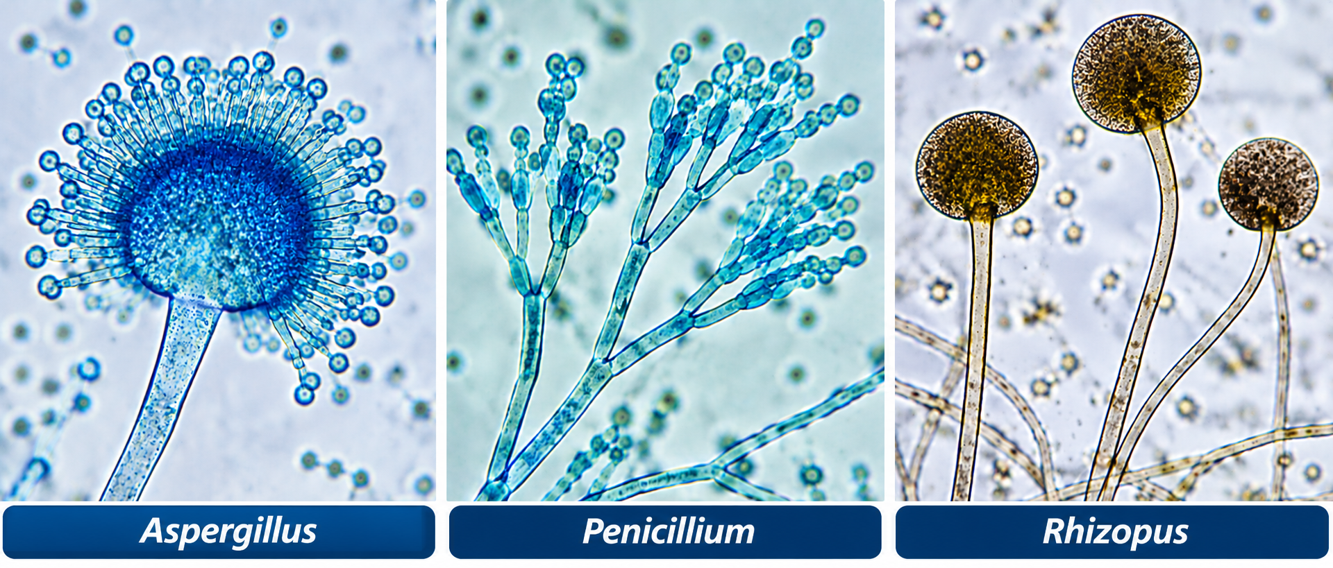
Different mold groups have recognizable microscopic characteristics. Here are several commonly studied examples.
Aspergillus Under a Microscope
Aspergillus is a widespread fungal genus found in soil, decaying vegetation, and various indoor and outdoor environments.
Under a compound microscope, characteristic features of many Aspergillus species include upright conidiophores ending in enlarged structures that support chains of conidia.
Depending on the species and preparation, the spore-bearing head may look rounded, columnlike, or radiating.
These structures are often examined at 100x to 400x magnification.
Penicillium Under a Microscope
Penicillium species are known for their distinctive branching reproductive structures.
Under magnification, the conidiophores can resemble tiny brushes, with chains of spores extending from their branches.
This brushlike arrangement is a useful characteristic when studying Penicillium , although further examination may be required for identification.
Rhizopus (Bread Mold) Under a Microscope
Rhizopus is commonly associated with mold growth on bread and other organic materials.
Microscopic characteristics can include:
- Broad, branching hyphae
- Upright sporangiophores
- Rounded sporangia containing spores
- Rootlike structures called rhizoids
In prepared specimens, the rounded sporangia can be particularly noticeable.
Rhizopus provides an example of how fungal reproductive structures differ from those of Aspergillus and Penicillium.
Cladosporium Under a Microscope
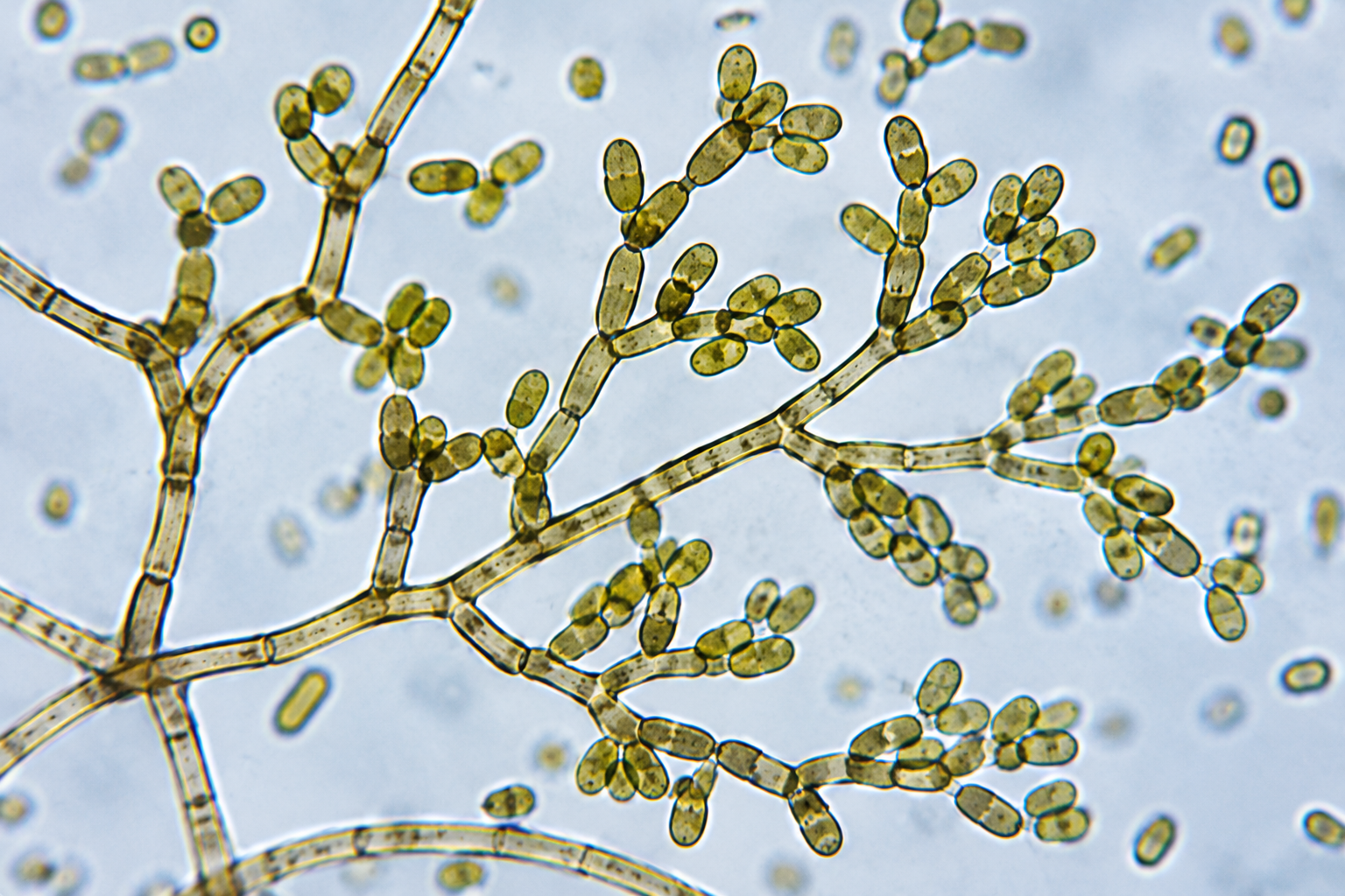
Cladosporium is another common fungal genus found in outdoor environments and sometimes indoors.
Its microscopic appearance may include pigmented, septate hyphae and branching chains of conidia.
The spores can vary in shape and may appear olive-brown or darker under transmitted light.
Microscopic examination can reveal these structural characteristics, but genus- or species-level identification requires appropriate expertise.
What Is the Difference Between Mold and Yeast Under a Microscope?
Mold and yeast are both fungi, but they often have different growth forms.
|
Feature |
Mold |
Yeast |
|
Typical structure |
Multicellular filaments |
Primarily single cells |
|
Appearance |
Branching hyphae and spore structures |
Round or oval cells |
|
Growth pattern |
Filamentous networks |
Budding or cell division |
|
Common microscopic features |
Mycelium, conidia, sporangia |
Budding cells and sometimes pseudohyphae |
Many yeasts reproduce by budding, which can make small daughter cells visible alongside larger parent cells.
Some fungi can exhibit both yeastlike and filamentous growth under different conditions, so this comparison is a general guide rather than an absolute distinction.
What Type of Microscope Is Best for Viewing Mold?
The best microscope depends on whether you're examining a prepared slide, studying fungal structures, or inspecting growth on a larger surface.
Compound Microscopes for Mold Spores and Hyphae
A compound light microscope is generally the best choice for examining mold spores, hyphae, and reproductive structures on prepared slides.
Look for features such as:
- 4x, 10x, and 40x objectives
- Adjustable transmitted illumination
- A mechanical stage for precise slide positioning
- Fine-focus controls for higher magnifications
- A 100x oil-immersion objective if your laboratory procedures require it
Stereo Microscopes for Surface Examination
A stereo microscope is useful for examining larger mold colonies, prepared materials, or surface growth at lower magnification.
Unlike compound microscopes, stereo microscopes provide a larger working distance and allow users to observe the three-dimensional appearance of specimens.
However, they generally cannot resolve the fine details of individual mold spores as effectively as a compound microscope.
Explore stereo microscopes for lower-magnification surface inspection applications.
Digital Microscopes for Documentation
For classroom demonstrations, research documentation, or sharing observations, a microscope equipped with a digital camera can capture images of fungal structures.
Digital imaging is particularly useful when comparing specimens or maintaining laboratory records.
Browse digital microscopes to explore microscope systems with imaging capabilities.
How to Observe Mold Under a Microscope Safely
Mold specimens should be handled carefully because disturbing fungal growth can release spores into the air.
For classroom and general educational use, commercially prepared, sealed microscope slides are the safest practical choice .
A basic observation process involves:
- Select a prepared mold slide appropriate for your microscope.
- Place the slide securely on the microscope stage.
- Begin with the lowest-power objective to locate the specimen.
- Adjust the illumination and focus until the fungal structures become visible.
- Increase magnification gradually to examine hyphae and reproductive structures.
- Record observations or capture images if a camera is available.
Avoid scraping mold from household surfaces, disturbing unknown colonies, or preparing slides from potentially hazardous material without suitable laboratory facilities and training.
Unknown mold samples may require containment and handling procedures that are not appropriate for home or classroom environments.
Can You Identify Black Mold Under a Microscope?
Microscopy can reveal characteristics of darkly pigmented fungi, but you cannot reliably identify toxic black mold based on color or appearance alone .
The phrase "black mold" is often associated with Stachybotrys chartarum , but numerous fungi can produce dark colonies or pigmented microscopic structures.
A visual examination cannot establish whether a specimen is Stachybotrys , whether it produces mycotoxins, or whether exposure presents a particular health risk.
Reliable identification may require professional laboratory analysis, including additional morphological examination or molecular methods.
If you suspect a significant indoor mold problem, consult an appropriately qualified professional rather than attempting to identify the material using a household microscope.
Why Scientists Study Mold Under a Microscope
Microscopy plays an important role in mycology, microbiology, environmental science, and industrial quality control.
Researchers and laboratory professionals use microscopes to:
- Examine fungal morphology and reproductive structures
- Compare prepared specimens
- Document fungal growth and development
- Support environmental and agricultural research
- Investigate fungal contamination in laboratory and industrial settings
- Teach students about fungal biology
In industrial and research applications, microscope selection may also depend on imaging requirements, sample preparation, illumination, and documentation needs.
Frequently Asked Questions About Mold Under a Microscope
Can You See Mold Spores with a Regular Microscope?
Yes. Many mold spores can be observed using a compound light microscope, particularly at 400x total magnification. The visibility of individual features depends on the species, specimen preparation, and optical quality of the microscope.
What Magnification Do You Need to See Mold?
A compound microscope with 100x to 400x total magnification is suitable for many mold observations. Lower magnifications can show larger fungal structures, while 400x is often useful for examining individual spores and hyphae.
What Color Is Mold Under a Microscope?
Mold can appear transparent, pale, brown, greenish, or darkly pigmented depending on the species, structures, lighting, and staining method. Colony color does not necessarily match the appearance of individual microscopic structures.
Can You See Mold With a 40x Microscope?
At 40x total magnification, you may see larger fungal structures or the overall arrangement of a prepared specimen. For detailed examination of spores and hyphae, a compound microscope offering 100x to 400x magnification is generally more useful.
Can You Tell if Mold Is Dangerous by Looking at It Under a Microscope?
No. Microscopic appearance alone cannot determine whether mold is toxic or whether it poses a health risk. Appropriate identification and risk assessment may require professional testing and evaluation.
Explore Mold and Fungal Structures with the Right Microscope
Viewing mold under a microscope reveals an intricate world of branching filaments, reproductive structures, and microscopic spores that cannot be appreciated with the naked eye.
For students and laboratories examining prepared fungal specimens, compound microscopes offer the magnification and optical control needed for detailed observations. Stereo microscopes are better suited to larger surface features, while digital imaging systems make it easier to document and share results.
Explore Microscope World's selection of
to find equipment for biology education, laboratory observation, and scientific research.


