
Comparison Microscopes: How They Work and What They're Used For
Nov 19th 2026
When forensic scientists examine two bullets, investigators compare hair samples, or laboratory technicians inspect similar materials, identifying small differences can be critical. A comparison microscope makes this process easier by allowing two specimens to be examined within a coordinated viewing system.
Unlike a conventional microscope that typically displays one specimen at a time, a comparison microscope is designed to help users examine two objects side by side, compare corresponding features, and identify similarities or differences.
In this guide, we'll explain how comparison microscopes work, their most common applications, and what to consider when choosing one for forensic or laboratory use.
What Is a Comparison Microscope?
A comparison microscope is a specialized optical instrument that allows users to view and compare two separate specimens through a combined viewing system. It is commonly used in forensic science, particularly for examining bullets, cartridge cases, toolmarks, and other physical evidence.
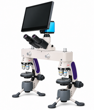
Traditional optical comparison microscopes typically use two microscope bodies connected by a comparison bridge. This arrangement allows the examiner to view images from both specimens simultaneously.
Depending on the system, the examiner may see:
- Two images positioned side by side
- A split field showing portions of both specimens
- Superimposed images for evaluating alignment and similarities
These viewing options help examiners evaluate corresponding details without repeatedly switching between specimens.
Comparison microscopes are particularly valuable when the differences being studied are small, subtle, or difficult to evaluate from memory.
How Does a Comparison Microscope Work?
A traditional comparison microscope uses two optical paths to examine separate specimens at the same time.
Each specimen is positioned beneath its own microscope objective. Light from both specimens travels through the optical system, where a comparison bridge combines the images into a shared viewing field.
The examiner can then adjust focus, illumination, magnification, and specimen position to evaluate specific features.
1. Two Specimens Are Positioned for Examination
The examiner places a reference specimen on one microscope stage and a questioned or unknown specimen on the other.
For example, a forensic examiner might compare a cartridge case recovered from a crime scene with a test-fired cartridge case.
2. Magnification and Lighting Are Adjusted
Both specimens are examined at appropriate magnifications.
Consistent illumination is important because differences in lighting can make similar surfaces appear different.
The examiner may adjust the lighting angle to make fine markings, scratches, or surface details easier to observe.
3. The Images Are Compared
The optical bridge allows the examiner to view both specimens in a coordinated field.
The examiner can move and rotate the specimens to compare corresponding features.
In a split-field view, the boundary between the two images can help reveal whether particular patterns appear to continue from one specimen to the other.
4. Findings Are Documented
Many modern comparison microscopes support digital cameras, image capture, and software.
These capabilities allow examiners to document observations, save comparison images, and share findings for review.
Importantly, a comparison microscope helps an examiner observe and document similarities and differences. It does not independently establish that two specimens came from the same source. Conclusions depend on the examination method, evidence quality, and applicable laboratory standards.
What Are Comparison Microscopes Used For?
Comparison microscopes are most closely associated with forensic science, but the underlying technique can also be useful in materials research and specialized inspection.
Forensic Ballistics and Firearm Examination
One of the best-known applications is the comparison of bullets and cartridge cases.
When a firearm is discharged, components of the firearm can leave microscopic markings on ammunition.
Examiners may compare:
- Striations on fired bullets
- Firing pin impressions
- Breechface impressions
- Extractor and ejector marks
- Other microscopic surface characteristics
A comparison microscope allows examiners to examine questioned evidence alongside test-fired samples.
These observations can contribute to a forensic examination, although the strength and interpretation of any association must be evaluated according to established procedures.
Toolmark Examination
Tools can leave microscopic impressions or striations on surfaces they contact.
Forensic examiners may use comparison microscopes to study marks associated with screwdrivers, cutting tools, or other instruments.
The goal is to evaluate the physical characteristics of questioned marks and compare them with marks produced under controlled conditions.
Hair and Fiber Examination
Comparison microscopy can also support the examination of hairs and textile fibers.
Examiners may compare characteristics such as:
- Color and pigmentation
- Diameter and structure
- Surface appearance
- Internal morphological features
- Fiber characteristics and visible differences
Microscopic hair comparisons cannot uniquely identify an individual. DNA analysis and other methods may be necessary when the investigation requires stronger source-specific evidence.
Document and Material Examination
Certain comparison systems may be used to examine printing characteristics, paper fibers, surface markings, and other physical details.
Applications depend on the microscope configuration, illumination options, and specimen dimensions.
Industrial Inspection and Quality Control
The same principle of comparing two specimens can be useful outside forensic laboratories.
For example, technicians may need to compare a manufactured component against a reference sample or examine differences between acceptable and defective materials.
For these applications, microscope selection should account for working distance, lighting, specimen size, and whether simultaneous optical comparison is necessary.
Comparison Microscope vs. Regular Microscope
The main difference is the ability to examine two separate specimens in a coordinated viewing field.
|
Feature |
Comparison Microscope |
Conventional Microscope |
|
Specimens viewed |
Two in a coordinated comparison |
Usually one at a time |
|
Optical configuration |
Two optical paths with comparison capability |
Single specimen imaging system |
|
Side-by-side viewing |
Core capability on traditional systems |
Usually requires separate images or software |
|
Common applications |
Forensics, evidence comparison, specialized inspection |
Biology, education, research, general inspection |
|
Digital imaging |
Available on many models |
Available on many models |
A conventional microscope can capture images of two specimens separately, and software may allow those images to be displayed side by side.
However, a traditional optical comparison microscope offers simultaneous viewing through an integrated optical system, which can be particularly useful when examining physical evidence.
What Magnification Does a Comparison Microscope Need?
There is no single magnification that works for every comparison microscopy application.
Lower magnifications are often useful for locating relevant surface features and examining larger specimens.
Higher magnifications allow closer inspection of fine markings and microscopic structures.
For example:
- 10x to 40x: Useful for examining larger objects, surface characteristics, and general evidence features.
- 40x to 100x: Useful for closer examination of selected markings and smaller structures.
- 100x to 400x: Useful for certain microscopic specimens and detailed observations, depending on the optical system.
The appropriate magnification depends on the specimen, objective lenses, working distance, and required resolution.
Forensic evidence such as bullets and cartridge cases often benefits from lower-power reflected-light examination, while prepared biological slides may require higher magnification and transmitted illumination.
How to Choose a Forensic Comparison Microscope
Before purchasing a comparison microscope, consider the types of specimens your laboratory examines and how observations will be documented.
Specimen Size and Working Distance
Larger evidence items require enough space beneath the objectives for positioning and adjustment.
If your work involves bullets, cartridge cases, or irregularly shaped objects, confirm that the microscope accommodates those specimens.
Illumination Options
Reflected illumination is important when examining opaque objects such as metal evidence.
Transmitted illumination may be useful for prepared slides and transparent specimens.
Some systems offer both illumination methods, increasing their versatility.
Comparison Viewing Modes
Consider whether the microscope provides side-by-side, split-field, or overlapping views.
Not every microscope marketed for forensic applications offers the same viewing capabilities.
Digital Imaging and Documentation
A camera can be useful for capturing comparison images, preparing reports, and sharing observations.
Depending on the system, imaging options may include a connected computer, tablet, or dedicated monitor.
Portability
Laboratories typically prioritize stable equipment and repeatable positioning.
However, portable or transportable systems may be useful for training, field examinations, and facilities where equipment needs to be moved.
Forensic Comparison Microscopes Available from Microscope World
Microscope World offers several comparison microscope configurations for forensic examination, education, and laboratory applications.
Swift M3-F Forensic Comparison Microscope
The Swift M3-F Forensic Comparison Microscope combines comparison viewing capabilities with a portable design.
Features include:
- Side-by-side and overlapping comparison views
- 10x, 40x, 100x, and 400x magnification
- Top and bottom LED illumination
- Trinocular head with a camera adapter
- Detachable tripod legs
This configuration may be useful for educational programs and laboratories that need to examine both opaque objects and prepared slides.
Swift M3-F-BTI2 Tablet Forensic Comparison Microscope
The Swift M3-F-BTI2 Tablet Forensic Comparison Microscope adds a tablet-based digital imaging system.
It provides comparison viewing capabilities alongside image capture and video recording features.
This makes it worth considering for laboratories and educational settings that need to document observations or display microscope images digitally.
HD WiFi Forensic Comparison Microscope
The HD WiFi Forensic Comparison Microscope offers comparison viewing with an HD camera, WiFi capabilities, and a 13-inch LCD monitor.
Its digital display may be useful for group demonstrations, image review, and laboratory documentation.
The system also supports both top and bottom illumination for examining different specimen types.
Frequently Asked Questions About Comparison Microscopes
What Is a Comparison Microscope Used For?
A comparison microscope is used to examine two specimens simultaneously and evaluate their similarities and differences. Common applications include forensic firearm examination, toolmark analysis, and certain types of trace evidence examination.
How Is a Comparison Microscope Different from a Compound Microscope?
A conventional compound microscope typically examines one specimen at a time using an objective and eyepiece system. A traditional comparison microscope combines images from two separate optical systems, allowing simultaneous comparison.
Why Are Comparison Microscopes Used in Forensics?
Comparison microscopes allow forensic examiners to study corresponding microscopic characteristics on questioned and reference specimens. This is particularly useful when examining firearm-related evidence, toolmarks, and certain trace materials.
Can a Comparison Microscope Identify a Bullet?
A comparison microscope can help an examiner evaluate microscopic markings on a bullet and compare them with markings on reference bullets. It cannot independently establish a definitive source identification. The examiner must interpret the observations using appropriate methods and standards.
Can Comparison Microscopes Capture Digital Images?
Yes. Many modern comparison microscopes support cameras, monitors, and software for capturing images and documenting observations. Available features vary by model.
Are Comparison Microscopes Only Used for Forensics?
No. Although forensic science is one of their primary applications, comparison microscopy can also support specialized material examination, research, education, and industrial inspection.
Find the Right Comparison Microscope for Your Application
Comparison microscopes make it possible to examine two specimens within a coordinated viewing system, helping users evaluate small physical characteristics that may be difficult to compare separately.
Whether you're examining forensic evidence, teaching microscopy techniques, or comparing specialized materials, the right system depends on your specimens, required magnification, illumination, and documentation needs.
Explore Microscope World's forensic comparison microscopes to find equipment suited to your laboratory or educational application.


