

What Does DNA Look Like Under a Microscope? Can You Actually See It?
Nov 2nd 2026
DNA is one of the most important molecules in every living organism. It carries the genetic instructions that determine how cells develop, function, and reproduce. But despite how often we see illustrations of DNA's famous double-helix structure, what does DNA actually look like under a microscope?
Individual DNA molecules are too small to see with a standard light microscope, even at 1000x magnification. However, scientists can observe DNA when it is tightly packaged into chromosomes, visualize extracted DNA as white, stringy material, and use specialized microscopy techniques to study individual DNA molecules.
What you see depends on the type of microscope, magnification, and how the DNA sample is prepared.
What Does DNA Actually Look Like?
DNA, or deoxyribonucleic acid, is a long molecule made of two strands twisted around each other. This structure is known as a double helix , often compared to a twisted ladder.
The sides of the ladder consist of sugar and phosphate molecules, while the rungs are formed by pairs of chemical bases: adenine (A), thymine (T), cytosine (C), and guanine (G).
Although illustrations commonly show DNA as a colorful spiral, actual DNA does not naturally appear blue, red, or green.
Its appearance depends on how it is observed:
- Individual DNA molecules: Extremely thin strands that require specialized imaging techniques to visualize.
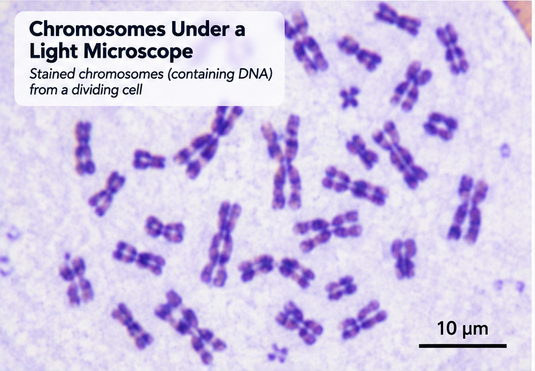
- Chromosomes: Condensed structures containing DNA and proteins that can appear as dark rods or X-shaped structures when stained.
- Extracted DNA: White, cloudy, or stringy material consisting of many DNA molecules collected together.
- Fluorescently labeled DNA: Brightly colored regions or structures against a darker background when viewed with appropriate fluorescence microscopy equipment.
The familiar double-helix illustrations are scientific models rather than what someone would see through an ordinary microscope.
Can You See DNA Under a Light Microscope?
No, a conventional light microscope cannot resolve individual DNA molecules. DNA's double helix measures approximately 2 nanometers in diameter, while a typical light microscope can resolve details only down to approximately 200 nanometers.
This means DNA is roughly 100 times thinner than the smallest details a conventional light microscope can distinguish.
Even a laboratory-grade compound microscope operating at 1000x magnification cannot reveal the individual strands of the DNA double helix.
However, a light microscope can reveal larger structures that contain DNA.
Viewing Chromosomes Under a Microscope
Chromosomes are structures made of DNA wrapped around proteins called histones. When cells prepare to divide, their chromosomes become tightly condensed, making them easier to observe.
With appropriate staining and sample preparation, chromosomes can appear as dark, elongated structures under a compound microscope.
Depending on the stage of cell division, they may look like:
- Short, thick rods
- Curved or threadlike structures
- X-shaped chromosomes consisting of two sister chromatids
The X shape is particularly recognizable during certain stages of cell division.
Chromosomes are often studied at magnifications of 400x to 1000x using a compound light microscope.
Important distinction: When you observe a chromosome, you are seeing DNA packaged together with proteins, not the individual DNA double helix.
What Does DNA Look Like Under an Electron Microscope?
Electron microscopes provide much greater resolving power than conventional light microscopes.
Instead of visible light, they use beams of electrons to examine extremely small structures.
With specialized preparation and imaging techniques, individual DNA molecules can appear as thin, threadlike structures.
Depending on the technique, DNA may look like:
- Long, narrow filaments
- Curved or tangled threads
- Loops and coiled structures
- Networks of overlapping strands
However, even electron microscopy does not automatically produce the familiar textbook image of a clearly defined DNA double helix.
Researchers use specialized techniques, including electron microscopy and atomic force microscopy, to investigate DNA's physical structure.
Can You See the DNA Double Helix?
The double-helix structure cannot be resolved with an ordinary light microscope.
Scientists have studied DNA's molecular structure using techniques such as X-ray diffraction, cryo-electron microscopy, and atomic force microscopy.
These approaches provide structural information at scales far smaller than conventional light microscopy can achieve.
The recognizable twisted-ladder model is based on scientific evidence about DNA's molecular arrangement, not simply a photograph taken through a classroom microscope.
What Magnification Do You Need to See DNA?
The magnification needed depends on whether you want to observe cells, chromosomes, or individual DNA molecules.
|
Magnification |
What You Can See |
|
40x |
Larger cells and tissue structures |
|
100x |
Groups of cells and some cellular details |
|
400x |
Cell nuclei and some stained chromosomes |
|
1000x |
More detailed views of stained chromosomes |
|
Electron or specialized nanoscale microscopy |
Individual DNA molecules with suitable preparation |
No amount of conventional light microscope magnification can reveal the individual DNA double helix.
This is because resolution, rather than magnification alone, determines how much detail a microscope can distinguish.
Increasing magnification beyond the instrument's resolving power simply produces a larger image without revealing additional structural detail.
For students and laboratory researchers interested in examining cells and chromosomes, a quality compound microscope is generally the appropriate starting point.
What Does Extracted DNA Look Like?
One of the easiest ways to observe DNA is through a simple extraction experiment using strawberries or other fruits.
Unlike the microscopic double helix, extracted DNA can form visible clumps when large numbers of DNA molecules collect together.
Extracted DNA typically looks like white, cloudy, stringy material. It may resemble mucus, thin cotton fibers, or small strands of wet thread.
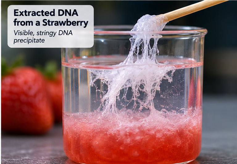
This material contains many DNA molecules grouped together rather than a single visible strand.
How to Extract DNA From a Strawberry
Strawberries are frequently used in classroom DNA experiments because they are easy to mash and contain substantial amounts of genetic material.
Materials needed:
- 2 to 3 fresh strawberries
- Water
- Dish soap
- Table salt
- A resealable plastic bag
- A coffee filter
- A clear container
- Chilled rubbing alcohol
- A wooden stick or toothpick
Step 1: Mash the strawberries
Place the strawberries in a resealable bag and crush them thoroughly to break apart the fruit tissue.
Step 2: Add the extraction solution
Mix approximately half a cup of water with two teaspoons of dish soap and half a teaspoon of salt.
Add a small amount of this solution to the mashed strawberries and mix gently.
The detergent helps disrupt cell membranes, releasing cellular contents, including DNA.
Step 3: Filter the mixture
Pour the mixture through a coffee filter into a clear container to remove larger pieces of fruit.
Step 4: Add chilled alcohol
Carefully pour cold rubbing alcohol down the side of the container to form a separate layer above the strawberry mixture.
DNA is poorly soluble in alcohol under these conditions, causing it to collect into visible material.
Step 5: Observe the DNA
Look for cloudy white strands forming near the boundary between the liquid layers.
You may be able to collect some of the material using a wooden stick.
The visible material is a crude DNA-rich precipitate that can also contain other cellular substances.
Safety note: Rubbing alcohol is flammable and should not be consumed. Children should perform this experiment with adult supervision.
Can You Put Extracted DNA Under a Microscope?
Yes, but the results may be different from what you expect.
Under a light microscope, extracted DNA may appear as translucent clumps, cloudy material, or tangled fibers.
You will not see individual DNA molecules or their double-helix structure.
The experiment is valuable because it demonstrates how DNA can be separated from cells and collected in quantities large enough to see.
What Type of Microscope Is Best for Studying DNA?
The best microscope depends on what you want to investigate.
Compound Light Microscopes
A compound microscope is ideal for observing cells, nuclei, and appropriately prepared chromosomes.
These microscopes commonly provide magnification between 40x and 1000x and are widely used in schools, universities, and biological laboratories.
They are useful for:
- Observing plant and animal cells
- Examining stained cell nuclei
- Studying chromosomes during cell division
- Teaching introductory genetics and cell biology
For additional information about how these instruments work, read our guide to compound microscopes .
Fluorescence Microscopes
Fluorescence microscopy allows researchers to identify and visualize DNA-containing structures using fluorescent dyes or labeled probes.
For example, certain fluorescent stains bind to DNA, causing nuclei or chromosomes to glow when illuminated with the appropriate excitation light.
Researchers use fluorescence microscopy to study chromosome organization, cell division, and the location of specific genetic sequences.
However, standard fluorescence microscopy does not resolve the individual DNA double helix.
Electron Microscopes and Specialized Instruments
For investigating individual DNA molecules and nanoscale structures, scientists use specialized instruments such as electron microscopes and atomic force microscopes.
These technologies are primarily used in advanced research settings and require specialized sample preparation.
Frequently Asked Questions About DNA Under a Microscope
Can you see DNA under a microscope at 1000x?
You cannot resolve individual DNA molecules at 1000x using a conventional light microscope. However, you can observe stained chromosomes containing DNA, provided the sample has been prepared correctly.
What color is DNA under a microscope?
DNA has no inherent visible color. Extracted DNA generally appears white or cloudy, while stained chromosomes may appear purple, blue, or another color depending on the stain. Fluorescently labeled DNA can appear brightly colored in fluorescence images.
Can you see DNA with a school microscope?
A school compound microscope can reveal cell nuclei and, with suitable prepared slides, chromosomes. It cannot show individual DNA molecules or the double-helix structure.
Can you see DNA with the naked eye?
Yes, when large quantities of DNA are extracted and precipitated together, they can form visible white, stringy material. Individual DNA molecules remain far too small to see without specialized instruments.
What is the difference between DNA and chromosomes?
DNA is the molecule that carries genetic information. Chromosomes are organized structures made of DNA and associated proteins. Chromosomes become especially visible under a microscope when they condense during cell division.
Can you see DNA in a cheek cell?
A standard light microscope can reveal the nucleus of a stained cheek cell, which contains DNA. However, the DNA molecules themselves cannot be individually resolved.
Explore the Microscopic World of Cells and Genetics
Although individual DNA molecules cannot be seen with a conventional light microscope, microscopy remains essential for studying genetics, cell biology, and chromosome structure.
From examining stained chromosomes to observing cell division, the right microscope allows students and researchers to explore the structures that contain our genetic information.
At Microscope World, we offer laboratory-grade microscopes for education, biological research, and scientific applications.
Whether you're equipping a classroom or working in a research laboratory, understanding what you want to observe is the first step toward selecting the right microscope.


