
Parts of a Compound Microscope: Labeled Diagram, Functions & Guide
Sep 16th 2026
A compound microscope uses a combination of lenses, illumination components and mechanical controls to produce a magnified view of specimens that are too small to see clearly with the naked eye. While designs vary between models, most compound microscopes share the same essential parts.
The main parts of a compound microscope include the eyepiece, objective lenses, revolving nosepiece, stage, condenser, iris diaphragm, illuminator, coarse and fine focus knobs, arm and base. Each part has a specific function in magnifying, illuminating, positioning or focusing the specimen.
Understanding these parts is useful whether you are learning to use a microscope for the first time, teaching microscopy in a classroom or choosing a microscope for laboratory work.
Labeled Parts of a Compound Microscope
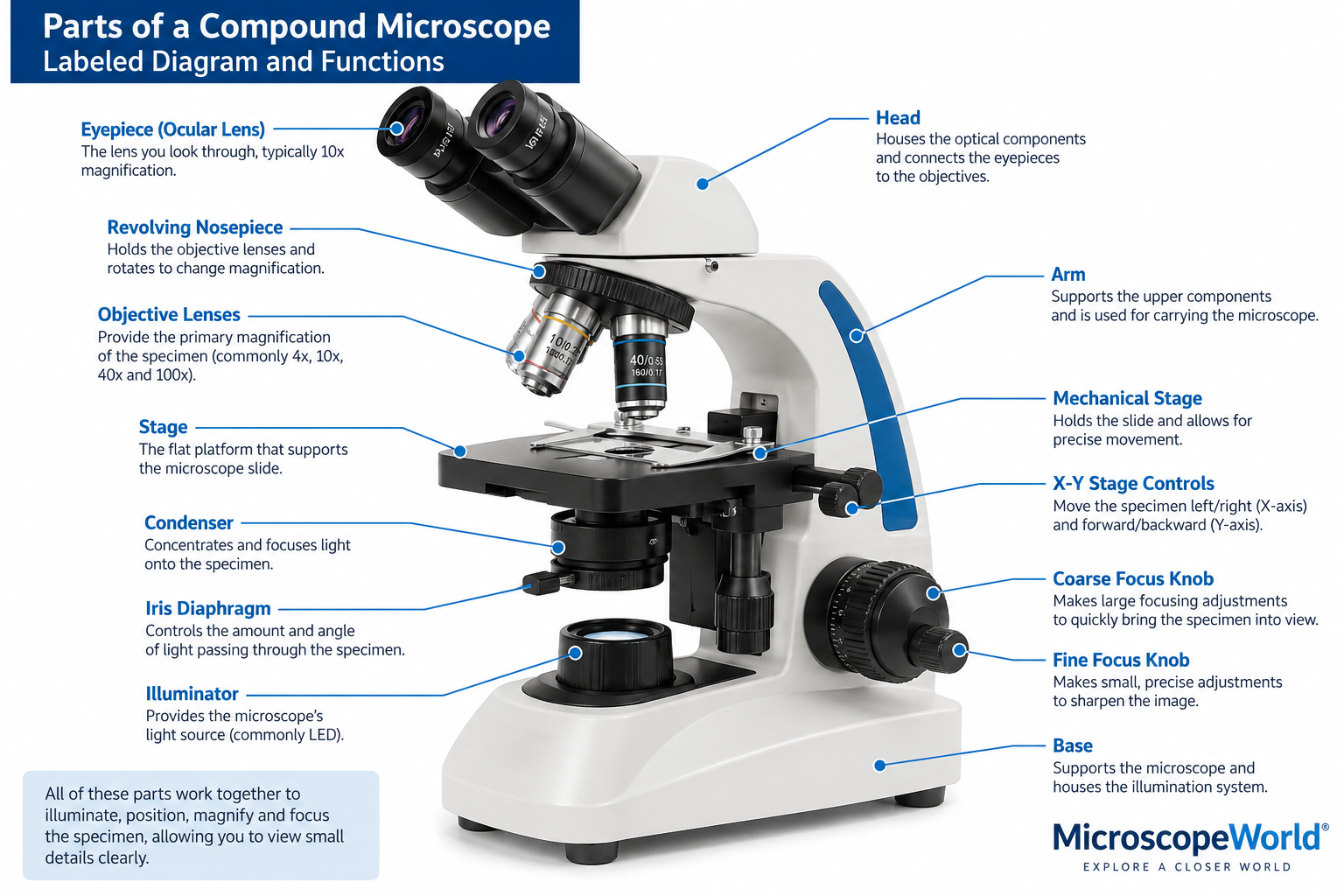
A labeled compound microscope diagram makes it easier to understand where each component is located and how the parts work together.
A typical compound microscope includes:
- Eyepiece or ocular lens
- Head or body
- Revolving nosepiece
- Objective lenses
- Stage
- Mechanical stage or stage clips
- X-Y stage controls
- Condenser
- Iris diaphragm
- Illuminator
- Coarse focus knob
- Fine focus knob
- Arm
- Base
Some microscopes may also include additional controls or specialized components depending on the model and intended application.
Compound Microscope Parts and Functions
Here is a quick reference to the major parts of a compound microscope and what they do.
|
Microscope Part |
Function |
|
Eyepiece/Ocular Lens |
Magnifies the image for viewing, commonly at 10x |
|
Objective Lenses |
Provide the primary magnification of the specimen |
|
Revolving Nosepiece |
Holds the objectives and rotates to change magnification |
|
Head/Body |
Houses optical components and connects the eyepiece to the objectives |
|
Stage |
Supports the microscope slide |
|
Mechanical Stage |
Holds and precisely moves the slide |
|
X-Y Stage Controls |
Move the specimen left/right and forward/backward |
|
Condenser |
Concentrates and focuses light onto the specimen |
|
Iris Diaphragm |
Controls the amount and angle of light passing through the specimen |
|
Illuminator |
Provides the microscope's light source |
|
Coarse Focus Knob |
Makes large focusing adjustments |
|
Fine Focus Knob |
Makes small, precise focusing adjustments |
|
Arm |
Supports the upper microscope components |
|
Base |
Supports the microscope and often contains the illumination system |
Optical Parts of a Compound Microscope
The optical system is responsible for producing and magnifying the image you see through the microscope.
Eyepiece or Ocular Lens
The eyepiece is the lens you look through at the top of the microscope. Most compound microscopes use a 10x eyepiece, although other magnifications may be available.
The eyepiece provides additional magnification to the image produced by the objective lens.
A monocular microscope has one eyepiece, a binocular microscope has two eyepieces, and a trinocular microscope typically has two viewing eyepieces plus an additional optical port for attaching a microscope camera.
Objective Lenses
The objective lenses are located close to the specimen and provide most of a compound microscope's magnification and resolving power.
Common objective magnifications include:
- 4x scanning objective
- 10x low-power objective
- 40x high-power objective
- 100x oil immersion objective
Not every microscope includes all four.
The objective you select determines how much of the specimen you can see, the level of detail visible and the working distance between the objective and specimen.
Higher magnification is not automatically better. The appropriate objective depends on the specimen and the level of detail you need to observe.
Revolving Nosepiece
The revolving nosepiece, sometimes called a turret, holds the objective lenses.
Rotating the nosepiece moves a different objective into the optical path, allowing you to switch magnification without changing lenses manually.
When changing objectives, rotate the nosepiece until the desired objective clicks securely into position.
Microscope Head
The head contains optical components that direct the image from the objective lenses toward the eyepieces.
Depending on the microscope, the head may be monocular, binocular or trinocular.
On many modern microscopes, the head can rotate to make viewing easier or allow multiple users to share the microscope.
Mechanical Parts of a Compound Microscope
Mechanical components hold the microscope together, support the specimen and allow the user to position and focus it.
Stage
The stage is the flat platform where the microscope slide is placed.
A hole or opening in the center of the stage allows light from the illuminator and condenser to pass through the specimen and into the objective lens.
Stage Clips
Basic microscopes may use stage clips to hold a slide in place.
The user moves the slide manually to examine different areas of the specimen.
Mechanical Stage
A mechanical stage provides greater control over specimen positioning.
Instead of moving the slide by hand, the user operates stage controls that move it precisely along two axes.
Mechanical stages are especially useful when working at higher magnifications, where even a small movement can shift the specimen significantly within the field of view.
X-Y Stage Controls
The X-Y controls move the slide in two directions.
One control moves the specimen left and right along the X-axis. The other moves it forward and backward along the Y-axis.
Together, these controls allow you to systematically examine different areas of a specimen.
Coarse Focus Knob
The coarse focus knob makes relatively large adjustments to the distance between the objective and specimen.
It is primarily used to bring the specimen into general focus at low magnification.
Because it produces large movements, coarse focus should generally not be used with high-power objectives when the objective is positioned very close to the slide.
Fine Focus Knob
The fine focus knob makes much smaller adjustments than the coarse focus control.
Once the specimen is approximately in focus, fine focus is used to sharpen the image and reveal finer details.
Fine focus becomes increasingly important as magnification increases.
Arm
The arm supports the upper portion of the microscope and connects it to the base.
It also provides a secure location for holding many microscope designs when moving them. When carrying a microscope, it is generally best to support the microscope with one hand at the arm and the other beneath the base.
Base
The base is the bottom portion of the microscope.
It provides stability and supports the microscope's weight. On modern compound microscopes, the base commonly houses the illuminator and portions of the electrical system.
Illumination Parts of a Compound Microscope
Magnification alone cannot produce a useful microscope image. The specimen must also be properly illuminated.
Several microscope components work together to control the light reaching the specimen.
Illuminator
The illuminator is the microscope's light source.
Many modern compound microscopes use LED illumination because LEDs provide bright, consistent light while producing relatively little heat and offering long operating life.
Some microscopes use other types of illumination depending on their design and application.
The illuminator directs light upward through the optical system toward the specimen.
Condenser
The condenser is located beneath the stage.
Its job is to collect and concentrate light from the illuminator onto the specimen.
Correct condenser positioning can have a major effect on image quality, especially when using higher-power objectives.
Iris Diaphragm
The iris diaphragm is generally part of or associated with the condenser assembly.
It adjusts the aperture of the illumination system, influencing the cone of light reaching the specimen. Adjusting the diaphragm affects image contrast, resolution and depth of field.
Closing the diaphragm can increase contrast, but closing it too far may reduce resolution. Opening it allows a wider cone of light to reach the objective.
Finding the proper setting is part of achieving a clear, well-balanced microscope image.
How the Parts of a Compound Microscope Work Together
The easiest way to understand a compound microscope is to follow the path that light takes through the instrument.
Illuminator → Condenser → Specimen → Objective Lens → Eyepiece → Eye
First, the illuminator produces light.
The condenser focuses that light onto the specimen positioned on the stage. The iris diaphragm helps control the illumination reaching the specimen.
Light passing through the specimen then enters the selected objective lens. The objective produces the primary magnified image.
That image travels through the microscope's optical system and reaches the eyepiece, which magnifies it further for viewing.
At the same time, the mechanical stage and focus controls allow the user to position the specimen and bring the image into sharp focus.
Every major component therefore contributes to one of four basic jobs:
Illuminate the specimen. Position the specimen. Magnify the image. Focus the image.
How Is Compound Microscope Magnification Calculated?
The total magnification of a compound microscope is calculated by multiplying the magnification of the eyepiece by the magnification of the objective lens.
Total Magnification = Eyepiece Magnification × Objective Magnification
For example, with a 10x eyepiece:
|
Objective |
Eyepiece |
Total Magnification |
|
4x |
10x |
40x |
|
10x |
10x |
100x |
|
40x |
10x |
400x |
|
100x |
10x |
1,000x |
This is why a microscope with a 10x eyepiece and 40x objective provides 400x total magnification.
However, magnification is only one aspect of microscope performance. Resolution, numerical aperture, illumination and optical quality determine how much useful detail can actually be observed.
Simply increasing magnification does not necessarily reveal additional detail.
How to Use the Main Compound Microscope Controls
For most specimens, begin with the lowest-power objective.
Place the prepared slide on the stage and secure it with the stage clips or mechanical stage. Position the area you want to examine over the stage opening.
Rotate the lowest-power objective, commonly 4x, into place.
Use the coarse focus control to bring the specimen into general focus. Adjust the illumination and diaphragm until the specimen is clearly visible.
Then use fine focus to sharpen the image.
Once the specimen is centered and focused, rotate to the next objective if additional magnification is needed. At higher magnifications, rely primarily on the fine focus control.
Starting at low magnification makes it easier to locate and center the specimen before examining smaller details.
Compound Microscope Parts That Are Commonly Confused
Some microscope components perform related functions, which can make their roles confusing for new users.
Condenser vs. Iris Diaphragm
The condenser concentrates and focuses light onto the specimen , while the iris diaphragm controls the aperture and cone of illumination .
They work together but perform different functions.
Coarse Focus vs. Fine Focus
The coarse focus knob makes large focusing adjustments and is typically used first at low power.
The fine focus knob makes small adjustments and is used to achieve a sharp image, particularly at higher magnifications.
Eyepiece vs. Objective Lens
Both magnify the specimen, but they operate at different stages of the optical system.
The objective lens provides the primary magnified image, while the eyepiece magnifies that image again for viewing.
Stage Clips vs. Mechanical Stage
Stage clips simply hold a microscope slide in place.
A mechanical stage both holds the slide and allows it to be moved precisely using X-Y controls.
Magnification vs. Resolution
Magnification describes how much larger an image appears.
Resolution describes the ability to distinguish two closely spaced points as separate details.
A microscope can produce a highly magnified image without providing additional useful detail if its optical system cannot resolve the specimen at that level.
Choosing a Compound Microscope
Understanding microscope parts also makes it easier to compare different compound microscopes.
For basic classroom use, features such as durable construction, straightforward focusing controls and LED illumination may be priorities.
College laboratories and more advanced educational environments may require higher-quality objectives, mechanical stages, binocular viewing and greater control over illumination.
Clinical, laboratory and research applications may require specialized optics, higher numerical aperture objectives, trinocular camera ports or techniques such as phase contrast, fluorescence or darkfield microscopy.
When comparing microscopes, consider how the complete optical and mechanical system supports the work you need to perform rather than looking at maximum magnification alone.
Microscope World offers compound microscopes for educational, laboratory, clinical and professional applications, along with microscope accessories, replacement components and imaging equipment.
Frequently Asked Questions About Compound Microscope Parts
What are the main parts of a compound microscope?
The main parts of a compound microscope include the eyepiece, objective lenses, revolving nosepiece, head, stage, condenser, iris diaphragm, illuminator, coarse and fine focus knobs, arm and base. Together, these components illuminate, position, magnify and focus the specimen.
What are the three main systems of a compound microscope?
The parts of a compound microscope can generally be grouped into optical, mechanical and illumination systems. The optical system magnifies the image, the mechanical system supports and positions the specimen and microscope components, and the illumination system provides and controls light.
What is the most important part of a compound microscope?
No single component works independently, but the objective lenses are particularly important because they provide the primary magnification and play a major role in determining resolution and image quality.
What does the objective lens do?
The objective lens collects light from the specimen and produces the primary magnified image. Compound microscopes usually have multiple objectives so users can switch between different levels of magnification.
What does the condenser do on a microscope?
The condenser collects and concentrates light from the illumination system onto the specimen. Proper condenser adjustment helps produce even illumination and improves image quality.
What does the diaphragm do on a compound microscope?
The iris diaphragm controls the aperture and cone of light used to illuminate the specimen. Adjusting it can change image contrast, resolution and depth of field.
What is the difference between coarse and fine focus?
Coarse focus makes larger adjustments and is generally used to initially locate the focus at low magnification. Fine focus makes smaller, more precise adjustments and is used to sharpen the image, especially at higher magnification.
Which parts of a compound microscope control magnification?
The objective lenses and eyepiece determine total magnification. Multiply the objective magnification by the eyepiece magnification to calculate total magnification.
Which microscope part controls the amount of light?
The microscope's illumination intensity control can adjust the brightness of the light source, while the iris diaphragm controls the aperture and cone of light passing through the illumination system.
Why should you start with the lowest-power objective?
A low-power objective provides a larger field of view and greater working distance, making it easier to locate, center and initially focus the specimen before moving to higher magnification.
What is the difference between a compound microscope and a stereo microscope?
A compound microscope is designed primarily for viewing small, thin specimens using transmitted light and relatively high magnification. A stereo microscope typically provides lower magnification, a larger working distance and a three-dimensional view that is useful for examining larger or solid specimens.
Understanding Your Compound Microscope
Learning the parts of a compound microscope makes the instrument much easier to use. Instead of viewing the microscope as a collection of knobs and lenses, you can understand how each component contributes to illumination, magnification, specimen positioning and focus.
Whether you are using a microscope in a classroom, clinical laboratory or research setting, proper use of the objectives, condenser, diaphragm, stage and focusing controls can make a significant difference in the quality of the image you observe.
When selecting a new microscope, these same components provide a useful framework for comparing models and choosing an instrument with the optical quality, controls and features required for your application.


