

Cheek Cells Under a Microscope: What They Look Like and How to View Them
Sep 8th 2026
Cheek cells are one of the easiest types of human cells to observe under a microscope. A simple sample collected from the inside of the cheek can reveal individual cells, their boundaries, cytoplasm and nuclei, providing a close-up look at the basic structures that make up the human body.
Under a microscope, human cheek cells typically appear as thin, flat, irregularly shaped cells with a clearly defined nucleus when properly stained. A compound microscope at 400x magnification usually provides the best view for identifying the major structures of a cheek cell.
Because cheek cells are easy to collect and require relatively simple equipment to observe, viewing them under a microscope is a classic biology experiment for students learning about cells, microscopy and the differences between animal and plant cells.
What Do Cheek Cells Look Like Under a Microscope?
Cheek cells under a microscope generally look like flat, irregular shapes with rounded edges. Unlike the rigid, brick-like appearance of many plant cells, cheek cells do not have a cell wall holding them in a fixed shape.
When a cheek cell sample is properly prepared and stained, you may be able to identify three primary structures:
- Cell membrane: The thin outer boundary surrounding each cell.
- Cytoplasm: The material filling the interior of the cell.
- Nucleus: A darker, usually rounded structure located within the cell.
The nucleus is often the most noticeable feature of a stained cheek cell.
Cheek cells may appear individually, but it is also common to see them overlapping or grouped together on a microscope slide. The appearance will vary depending on how the sample was collected, how much material was placed on the slide, the stain used and the microscope's magnification.
What Type of Cells Are Cheek Cells?
The cells collected from the inside of your cheek are squamous epithelial cells .
Epithelial tissue covers body surfaces and lines many internal structures. The epithelial cells inside the mouth create a protective barrier between underlying tissue and the environment inside the oral cavity.
"Squamous" refers to their relatively thin and flattened shape.
Cells are continually shed from the lining of the mouth and replaced by new cells. This is one reason a gentle swab of the inside of the cheek can collect enough cells for observation without needing to scrape aggressively.
Like other human cells, cheek cells are animal cells and eukaryotic cells . Eukaryotic cells contain a nucleus enclosed by a membrane.
What Magnification Do You Need to See Cheek Cells?
You can see groups of cheek cells at relatively low magnification, but 400x magnification is typically the best level for clearly observing individual cheek cells and their nuclei in a classroom microscope experiment.
Here's what you can generally expect at different magnifications:
|
Total Magnification |
What You May See |
|
40x |
The overall sample area and larger groups of stained material |
|
100x |
Groups of cheek cells and some individual cell shapes |
|
400x |
Individual cells, cell boundaries and stained nuclei |
|
1000x |
A more enlarged view, although usually unnecessary for a basic cheek cell experiment |
A standard compound microscope equipped with 4x, 10x and 40x objectives and a 10x eyepiece provides total magnifications of 40x, 100x and 400x.
For most students, 400x provides plenty of magnification to study cheek cell structure .
Higher magnification isn't automatically better. As magnification increases, the field of view becomes smaller and focusing becomes more difficult. The amount of useful detail you can see also depends on the microscope's optical resolution, not simply the magnification number.
What Type of Microscope Is Used to View Cheek Cells?
A compound light microscope is the best type of microscope for viewing cheek cells.
Compound microscopes are designed to examine thin, transparent specimens mounted on microscope slides. Light passes through the specimen from below and travels through the objective lens to produce a magnified image.
This makes them well suited for viewing biological specimens such as cells, tissue sections and microorganisms.
A typical student or classroom compound microscope capable of reaching 400x magnification is sufficient for a basic cheek cell experiment.
A stereo microscope, sometimes called a dissecting microscope, is generally not the best choice for cheek cells. Stereo microscopes operate at lower magnifications and are better suited for observing larger, three-dimensional objects such as insects, rocks, plants and manufactured components.
If you're choosing equipment for biology experiments like this one, Microscope World offers a range of compound microscopes and student microscopes designed for viewing prepared slides and biological specimens.
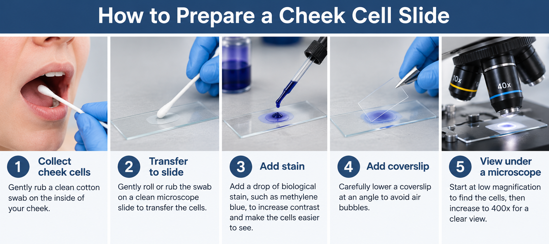
How to Prepare a Cheek Cell Microscope Slide
Preparing a cheek cell slide is relatively simple, but getting a thin, well-stained sample makes the cells much easier to observe.
What You'll Need
Gather the following materials:
- Compound microscope
- Clean microscope slide
- Coverslip
- Clean cotton swab
- Appropriate biological stain
- Dropper
- Water or saline, depending on the procedure
- Gloves and appropriate cleanup materials
Students should follow their instructor's laboratory procedures and appropriate safety practices when collecting, staining and disposing of biological samples.
1. Collect the Cheek Cells
Using a clean cotton swab, gently rub the inside surface of the cheek.
You do not need to scrape hard. A gentle swabbing action should collect enough loose epithelial cells for observation.
2. Transfer the Cells to the Slide
Gently rub or roll the swab across the center of a clean microscope slide to transfer the collected cells.
A thin sample is preferable. Too much material can create overlapping cells that are difficult to distinguish.
3. Add the Stain
Add the stain according to the procedure or stain manufacturer's instructions.
Cheek cells are nearly transparent, so staining increases contrast and makes structures such as the nucleus easier to distinguish.
4. Add a Coverslip
Carefully lower a coverslip over the specimen.
Lowering it gradually at an angle can help reduce trapped air bubbles, which can interfere with viewing.
5. Start at Low Magnification
Place the slide on the microscope stage and begin with the lowest-power objective.
Starting at low magnification provides a wider field of view, making it much easier to locate the sample.
6. Find and Focus on the Cells
Bring the sample into focus and look for areas containing separated cells rather than dense clumps.
Once you have located a good area, center it in the field of view.
7. Increase to 400x Magnification
Move progressively through the microscope's objective lenses until you reach 400x total magnification.
At 400x, individual cheek cells and their stained nuclei should become much easier to identify.
When using higher-power objectives, use the fine focus control as appropriate for your microscope rather than making large focus adjustments.
Why Are Cheek Cells Stained Before Viewing?
Cheek cells are largely transparent when viewed with an ordinary brightfield microscope. Without sufficient contrast, their internal structures can be difficult to distinguish from the surrounding background.
Staining cheek cells increases contrast, making structures such as the nucleus easier to see under a light microscope.
The stain interacts differently with cellular components, causing some structures to appear darker than others.
In a typical stained cheek cell, the nucleus will often appear as a dark spot or oval within the lighter body of the cell.
Staining is not unique to cheek cells. Scientists use a variety of staining techniques throughout microscopy to make otherwise transparent biological structures easier to identify and study.
Parts of a Cheek Cell You Can See Under a Microscope
A classroom light microscope cannot reveal every structure inside a human cell. Many cellular components are simply too small or lack enough contrast to be resolved using basic brightfield microscopy.
However, several major features can be identified.
Cell Membrane
The cell membrane forms the outer boundary of the cheek cell.
Animal cells do not have the rigid cell walls found in plant cells. As a result, cheek cells tend to have more irregular and rounded outlines.
Cytoplasm
The cytoplasm fills much of the space between the cell membrane and nucleus.
Under a standard classroom microscope, it may appear as a relatively light or translucent region surrounding the nucleus.
Nucleus
The nucleus is usually the easiest internal structure to identify in a properly stained cheek cell.
It often appears as a darker round or oval area within the cell.
The nucleus contains most of the cell's genetic material and helps regulate many cellular activities.
What About Mitochondria and Other Organelles?
Diagrams of animal cells commonly show mitochondria, the Golgi apparatus, endoplasmic reticulum, ribosomes and other organelles.
Don't expect to see all of these structures during a basic cheek cell experiment.
Many organelles are too small to be clearly resolved with an ordinary classroom light microscope or require specialized staining and imaging techniques. A diagram of an animal cell therefore contains considerably more detail than you will normally see when looking at actual cheek cells through a basic compound microscope.
Cheek Cells vs. Onion Cells Under a Microscope
Cheek cells and onion epidermal cells are frequently used together in biology classes because they provide a simple way to compare animal and plant cells.
The differences can be surprisingly obvious under a microscope.
|
Feature |
Human Cheek Cells |
Onion Epidermal Cells |
|
Cell type |
Animal cell |
Plant cell |
|
Typical shape |
Flat and irregular |
Rectangular or brick-like |
|
Cell membrane |
Yes |
Yes |
|
Cell wall |
No |
Yes |
|
Nucleus |
Visible with appropriate staining |
May be visible with appropriate preparation and staining |
|
Chloroplasts |
No |
Generally absent from onion bulb epidermal cells |
|
Arrangement |
May be scattered or overlapping |
Often arranged in an orderly pattern |
The most noticeable difference is usually their shape.
Onion cells have rigid cell walls that create clearly defined, rectangular boundaries. Human cheek cells lack a cell wall, giving them a softer and more irregular appearance.
Why Don't Onion Bulb Cells Have Visible Chloroplasts?
Students are often taught that plant cells contain chloroplasts, so viewing onion cells can cause some confusion.
The fleshy part of an onion bulb grows underground and is primarily used for food storage rather than photosynthesis. Its epidermal cells therefore generally lack the prominent chloroplasts you might see in photosynthetic tissues from green leaves.
Looking at both cheek and onion cells is a useful reminder that real cells don't always look exactly like simplified textbook diagrams.
Common Problems When Viewing Cheek Cells
If you're having trouble finding or identifying cheek cells, the problem is often related to slide preparation or microscope setup rather than the microscope itself.
I Can't Find Any Cells
Return to the lowest magnification and scan the slide slowly. Make sure you collected enough material and transferred it to the slide.
Everything Looks Like a Dark Clump
There may be too many cells or too much stain on the slide. A thinner sample can make individual cells easier to distinguish.
I Can See Cells, But Not the Nucleus
The sample may not have enough contrast. Proper staining and careful fine focusing can make the nucleus more visible.
I See Large Perfect Circles
You may be looking at air bubbles rather than cells. Air bubbles often have thick, dark outlines and a much more perfectly circular appearance.
The Image Becomes Blurry When I Increase Magnification
Before switching objectives, center the cells in your field of view and get the image sharply focused. After moving to a higher-power objective, make small adjustments using fine focus.
Also remember that increasing magnification cannot compensate for poor resolution or a poorly prepared sample.
What Can We Learn from Looking at Cheek Cells?
Viewing cheek cells is a simple experiment, but it demonstrates several important concepts in biology and microscopy.
Students can directly observe that the human body is made of individual cells rather than simply looking at an illustration in a textbook. They can also identify some of the major features shared by eukaryotic cells and compare real animal cells with plant cells.
The experiment also teaches fundamental microscopy skills, including:
- Preparing a wet-mount slide
- Using stains to increase contrast
- Starting observations at low magnification
- Centering a specimen before increasing magnification
- Using coarse and fine focus controls appropriately
- Recognizing artifacts such as air bubbles
- Comparing cell structures and shapes
These same basic skills form the foundation for more advanced microscopy in biology, medicine and scientific research.
Frequently Asked Questions About Cheek Cells Under a Microscope
What do cheek cells look like under a microscope?
Human cheek cells generally appear as thin, flat and irregularly shaped cells. When properly stained, the cell boundary and a darker nucleus can usually be distinguished under a compound microscope.
What magnification is best for viewing cheek cells?
Around 400x total magnification is typically ideal for viewing individual cheek cells and identifying their nuclei. Lower magnifications such as 40x and 100x are useful for locating the specimen before moving to higher power.
Can you see cheek cells at 40x magnification?
Groups of cells and stained material may be visible at 40x, but individual cellular structures are much easier to distinguish at higher magnification. Starting at 40x is useful for locating the sample.
Can you see the nucleus in a cheek cell?
Yes. The nucleus is typically visible in a properly prepared and stained cheek cell when viewed with a compound light microscope. It usually appears darker than the surrounding cytoplasm.
Why are cheek cells stained?
Cheek cells are naturally transparent. Staining increases contrast between cellular structures, making the cell and especially its nucleus easier to see.
What type of microscope is best for cheek cells?
A compound light microscope is best for viewing cheek cells because it provides the magnification and transmitted illumination needed to observe thin biological specimens on slides.
Why don't cheek cells have a cell wall?
Cheek cells are animal cells. Animal cells have flexible cell membranes but do not have the rigid cell walls characteristic of plant cells.
Are cheek cells living cells?
Cells collected from the cheek originate from living epithelial tissue, although once they have been removed, mounted, stained and prepared for conventional microscope observation, they should not be assumed to remain living.
What's the difference between cheek cells and onion cells?
Cheek cells are animal cells and tend to have irregular shapes without cell walls. Onion epidermal cells are plant cells with rigid cell walls that give them a more rectangular, organized appearance.
Can you see cheek cells without staining?
Cheek cells can be present and observable without staining, but their transparency makes them much harder to distinguish with a standard brightfield microscope. Staining increases contrast and makes the nucleus and cell boundaries easier to identify.
Explore the Microscopic World Around You
Cheek cells are an excellent example of how a microscope can reveal structures that are completely invisible to the naked eye. With a properly prepared slide and a compound microscope capable of approximately 400x magnification, you can observe individual human cells and identify features such as the cell membrane, cytoplasm and nucleus.
It's also an accessible starting point for learning how magnification, staining, contrast and specimen preparation affect what you can actually see through a microscope.
For classrooms, students and anyone interested in exploring cells and other biological specimens, Microscope World offers compound microscopes, student microscopes , prepared slides and microscopy accessories for educational and laboratory applications.


