The images found below were all captured with microscope digital cameras that we offer. All images shown were compressed to load faster on your computer, but are otherwise untouched photos.

Images taken using a digital camera on a ML7100 Metallurgical Microscope

Aluminum 100x
Aluminum 200x
Aluminum 400x
Anodized aluminum, 100x
Anodized aluminum, 200x
Anodized aluminum, 400x
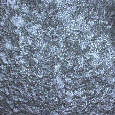
The anodized aluminum piece at the left is the same that was placed under the metallurgical microscope and photographed with the DK3000 digital camera. (See images above).  
     
microscope image, steel, 100x
microscope image, steel, 200x
microscope image, steel, 400x
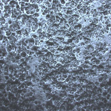
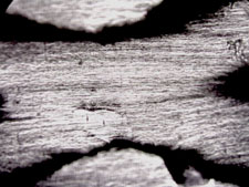
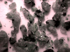
Scratched stainless steel, 100x
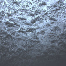
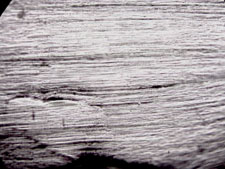
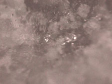
Scratched stainless steel, 200x
Scratched stainless steel, 400x
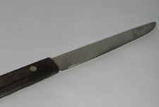
This old kitchen knife was used to capture the scratched stainless steel photos above. The metallurgical microscope provided both good magnification and illumination to capture images with the DK3000 digital camera.  
     
microscope image - plastic, 100x
microscope image - plastic, 200x
microscope image - plastic, 400x
Plastic strap, 100x
Plastic strap, 200x
Plastic strap, 400x
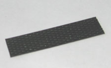
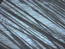
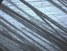
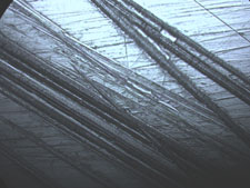
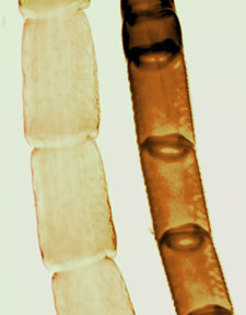
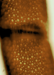
This plastic strap was originally used to keep the outside of a packing box closed. It was used under the metallurgical microscope to capture the images above.  

Images taken using a digital microscope camera on a student biological microscope.

Microscope image of aphid

The image at the left is an aphid.

This image was captured at 100x magnification.

   
Antennae 100x

Image at the left are two different insect antenna. This image was captured at 100x magnification.


Image at right is the exact same antenna as that in the image on the left, but the image was captured at 400x magnification.

Antennae 400x
     
Frog blood 400x

Image at the left is frog's blood, notice the small dark dots in the center - these are the nuclei. This image was captured at 400x magnification.


Image at the right is the same microscope slide with frog's blood, but was captured at 1000x magnification. Now the nuclei of the blood cells are clearly visible.

Frog Blood 1000x

Images taken using a digital stereo microscope.

Microscope image, carpet fiber, 10x

The image at the left is carpet fiber. This image was captured at 10x magnification.


Image at the right is the same carpet fiber, but this image was captured at 40x magnification.

Microscope image, carpet fiber, 40x
Microscope image of granite, 10x

The image at the left is granite. This image was captured at 10x magnification.


Image at the right is the same granite, but this image was captured at 40x magnification.

Microscope image of granite, 40x

Image taken using a digital stereo microscope.

Microscope image, PCI card, 10x

Image at the left is a PCI card.

This image was captured at 10x magnificaiton.