

Daphnia under the Microscope
Sep 26th 2017
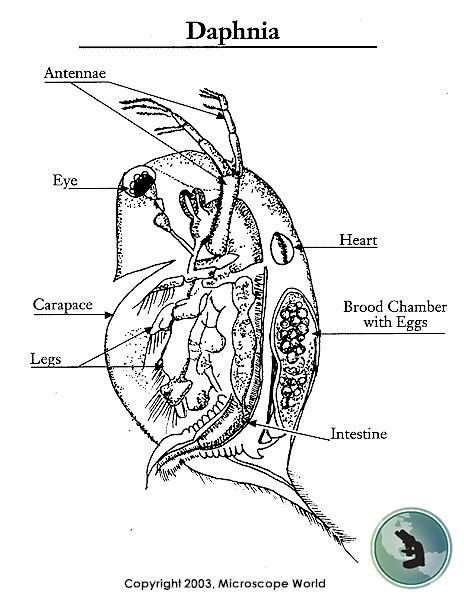
What Does Daphnia Look Like Under The Microscope?
Daphnia are a genus of small planktonic crustaceans that are commonly referred to as water fleas. Daphnia are called water fleas because their swimming style resembles the movement of fleas. Typically 0.2-5mm in length, Daphnia live in a variety of aquatic environments including acidic swamps, ponds, streams, freshwater lakes and rivers.
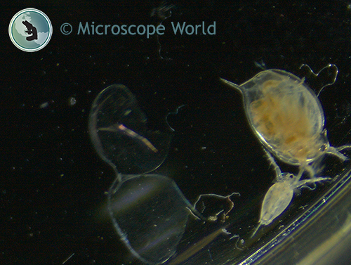
In this image there are two Daphnia captured under the Zeiss Stemi 508 stereo microscope. The smaller Daphnia on the bottom shows a ventral view of an adult female. Notice the antenna, single black compound eye in the center and the apical spine protruding from the end of the body.
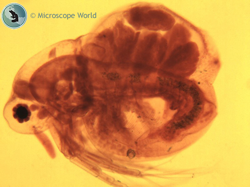
Below is an image of Daphnia that was captured using brightfield microscopy with a compound microscope at 100x magnification. The National Optical DC5-169 microscope was used to capture the image.
Because Daphnia are larger in size (1-5mm) and often in a petri dish, they are typically viewed under a stereo dissecting microscope.  Transmitted light and darkfield illumination were used in the image shown at right to capture this Daphnia. This image was captured with a darkfield stereo microscope.
To learn more about Daphnia visit NIH's in-depth page on Daphnia biology.
If you have questions regarding the correct microscope for your application contact Microscope World.


