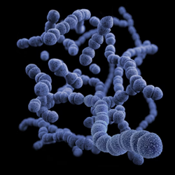
Tips for Viewing Bacteria Under a Microscope
Jun 18th 2023
Exploring the Microbial Universe: Tips for Viewing Bacteria Under a Microscope
 Bacteria, the tiny microorganisms that populate our world, hold significant importance in fields such as microbiology, medicine, and environmental science. Viewing bacteria under a microscope allows us to unlock their fascinating structures, behaviors, and interactions. However, obtaining clear and detailed observations of bacteria requires careful preparation and techniques. In this article, we will provide valuable tips to enhance your experience and proficiency in viewing bacteria under a microscope.
Preparing Microscopic Slides
Proper slide preparation is essential for successful bacterial observation. Follow these steps:
a. Start with a clean glass slide and cover slip to prevent contamination.
b. Sterilize the slide by passing it through a flame or using a disinfectant solution.
c. Place a small drop of the bacterial sample on the slide, ensuring even distribution.
d. Gently place the cover slip on top, avoiding air bubbles. Press down gently to flatten the sample.
Staining Techniques
Staining enhances the visibility of bacteria by adding contrast and color. You can learn about commonly used microscope stains here. Consider using the following staining techniques:
a. Gram Staining: This technique differentiates bacteria into Gram-positive (purple) and Gram-negative (pink) based on their cell wall characteristics.
b. Acid-Fast Staining: Useful for identifying bacteria with waxy cell walls, such as Mycobacterium tuberculosis, the causative agent of tuberculosis.
c. Differential Stains: Techniques like the Ziehl-Neelsen or Spore Stain allow for specific identification of particular bacteria or structures.
Adjusting Microscope Settings
To optimize your bacterial observations, ensure the microscope is properly set up:
a. Start with the lowest magnification objective lens to locate and center the bacteria.
b. Gradually increase the magnification to obtain finer details.
c. Adjust the focus using the coarse and fine adjustment knobs to achieve a clear image.
d. Modify the condenser and diaphragm settings to control the intensity and quality of the light.
Lighting Techniques
Appropriate lighting techniques are vital for enhancing bacterial visibility:
a. Use brightfield illumination to clearly view the bacteria.
b. Adjust the light intensity to avoid overexposure or underexposure of the specimen.
c. Utilize phase contrast microscopy to visualize transparent or unstained bacteria.
Observation and Recording
Follow these tips to optimize your bacterial observations:
a. Scan the entire slide to locate areas with a concentration of bacteria.
b. Focus on different fields of view to examine the diversity and distribution of bacteria.
c. Take time to adjust the focus and lighting for each area of interest.
d. Capture clear and well-lit images or videos to document your findings and aid further analysis.
Maintenance and Hygiene
Maintaining a clean and sterile environment is crucial for accurate bacterial observations:
a. Regularly clean and disinfect your microscope to prevent contamination.
b. Use sterile techniques when handling bacterial samples and slides.
c. Store slides and cover slips in clean, dust-free containers.
d. Properly dispose of bacterial waste and disinfect surfaces after use.
Viewing bacteria under a microscope offers a captivating journey into the microbial world. By following these tips for slide preparation, staining, microscope settings, lighting techniques, and observation, you can enhance your ability to observe bacteria in detail. Remember to maintain a sterile environment and record your findings accurately. With practice and attention to detail, you can unlock the hidden secrets of bacteria, contributing to advancements in various scientific disciplines.
Types of Bacteria To View Under A Microscope?
There are numerous types of bacteria that can be viewed under a microscope. Here are some commonly studied and observed bacteria:
Escherichia coli (E. coli): This bacterium is commonly found in the lower intestine of humans and other warm-blooded animals. It is used extensively in biological research and is a model organism for studying bacterial genetics and physiology.
Bacillus subtilis: This bacterium is commonly found in soil and the gastrointestinal tract of animals. It has been widely studied for its ability to form spores and its use as a model organism for various cellular processes.
Staphylococcus aureus: This bacterium is a common cause of skin infections and is often found in the nasal passages of humans. It is known for its golden-colored colonies and is an important pathogen in healthcare settings.
Streptococcus pneumoniae: This bacterium is a major cause of pneumonia, meningitis, and other respiratory infections. It can be observed under the microscope in chains or pairs (diplococci).
Salmonella enterica: This bacterium is a common cause of foodborne illnesses, such as salmonellosis. It can be observed as rod-shaped cells (bacilli) under the microscope.
Mycobacterium tuberculosis: This bacterium causes tuberculosis (TB), a contagious airborne disease. It appears as slender, rod-shaped cells and is often stained with special dyes for better visualization.
Vibrio cholerae: This bacterium is responsible for cholera, a severe diarrheal disease. It can be seen as curved or comma-shaped cells (vibrioids) under the microscope.
Lactobacillus acidophilus: This bacterium is a common probiotic found in the human gastrointestinal tract. It is used commercially in the production of yogurt and other fermented foods. It appears as rod-shaped cells under the microscope.
These are just a few examples of bacteria that can be viewed under a microscope. There are many other types of bacteria with diverse shapes, sizes, and characteristics that can be studied using microscopic techniques.
Frequently Asked Questions
How Do You View Bacteria Under a Microscope?
To view bacteria under a microscope, several steps need to be followed. Firstly, a clean microscope slide is prepared by removing any contaminants using a lint-free cloth or lens paper. A small drop of water or a bacterial growth medium is placed in the center of the slide.
Next, a bacterial sample is obtained from a culture plate, liquid culture, or other sources. Using a sterile loop or inoculating needle, a small amount of the sample is spread onto the water or growth medium drop on the slide. Care is taken to ensure the sample is evenly distributed and not too thick. The slide is then allowed to air dry completely.
Staining the bacterial sample is an optional step that can enhance contrast and visualization. Different staining methods, such as Gram staining or simple stains like methylene blue or crystal violet, can be used based on the specific requirements. The staining protocol is followed accordingly.
After staining (if applicable), a coverslip is placed over the sample, avoiding any air bubbles. Gently pressing down ensures proper adherence of the coverslip to the slide.
The compound light microscope is then turned on, and the light intensity is adjusted to an appropriate level. Starting with the lowest magnification objective (typically 10x or 20x), the slide is positioned on the microscope stage.
Observation begins by looking through the eyepiece and slowly adjusting the focus knobs to bring the bacteria into sharp focus. The slide can be moved around to explore different areas. If more detailed observation is required, higher power objectives (such as 40x or 100x) can be used.
During observation, attention is paid to the bacteria's shape, arrangement, size, and any other pertinent characteristics. These observations can be documented by sketching or capturing photographs through the microscope.
Throughout the process, it is important to handle all materials with care to maintain a sterile environment and prevent contamination. Following the specific instructions provided with the microscope ensures proper usage and maintenance.
What is the Best Way to Observe Bacteria?
The best way to observe bacteria is by using a compound light microscope. This type of microscope provides the necessary magnification and resolution to visualize bacteria. To ensure accurate observations, it is important to follow proper techniques.
Firstly, preparing a microscope slide is essential. A clean slide is used, and a small drop of water or bacterial growth medium is placed on it. A bacterial sample is obtained from a culture plate or liquid culture, and a small amount is spread onto the drop. The slide is allowed to air dry completely.
Staining the bacterial sample is often recommended for better visualization. Various staining methods can be used, such as Gram staining or simple stains like methylene blue. Staining helps to highlight the bacteria's characteristics and provides contrast against the background.
Once the slide is prepared, it is placed on the microscope stage. Starting with a low magnification objective, such as 10x or 20x, the bacteria are located by adjusting the focus knobs. Slow and careful movements ensure that the bacteria come into clear focus.
For more detailed observation, higher magnification objectives like 40x or 100x can be used. The slide can be moved to different areas to explore various regions of the bacterial sample.
During observation, attention should be given to the bacteria's shape, arrangement, and any other observable features. Detailed notes, sketches, or photographs can be taken to document the findings accurately.
Maintaining a sterile environment throughout the process is crucial to prevent contamination and obtain reliable observations. Following the specific instructions provided with the microscope and adhering to proper laboratory practices ensure optimal results.
Overall, using a compound light microscope, preparing the slide properly, and employing staining techniques when necessary provide the best way to observe bacteria and study their characteristics effectively.
What Magnification is Used to View Bacteria Under a Microscope?
To view bacteria under a microscope, various magnifications are typically used. The specific magnification depends on the size and level of detail required for observation. The most commonly used magnifications for viewing bacteria are:
Low magnification (10x to 20x): This magnification is useful for locating and surveying the bacterial sample on the microscope slide. It provides an initial overview of the distribution and arrangement of the bacteria.
Medium magnification (40x to 100x): This range of magnification allows for more detailed observation of bacterial morphology, such as shape, size, and cellular structures. It provides clearer visualization of individual bacteria and their characteristics.
High magnification (oil immersion, typically 100x to 1000x): High magnification objectives are used for finer examination of bacteria. Oil immersion microscopy is often employed at this level to maximize resolution. It involves placing a drop of immersion oil between the slide and the microscope objective to reduce light refraction and improve image clarity. This magnification is crucial for studying smaller bacterial structures, such as cell walls, flagella, and pili.
It's important to note that the maximum useful magnification of a compound light microscope is typically around 1000x. Beyond this, the resolution becomes limited by the wavelength of visible light, and electron microscopy is usually required for higher magnifications.
Choosing the appropriate magnification depends on the specific objectives of the study and the characteristics of the bacteria being observed. It may involve using different magnifications sequentially to obtain a comprehensive understanding of the bacterial sample.
How to Make Your Bacteria More Visible
To make bacteria more visible under a microscope, there are several techniques you can employ:
Staining: Staining is a common method used to improve the contrast and visibility of bacteria. Different staining techniques, such as Gram staining, acid-fast staining, or simple stains like methylene blue or crystal violet, can be used to highlight bacterial structures and make them more visible. Staining techniques involve applying specific dyes or chemicals to the bacterial sample, which adhere to different components of the bacteria, aiding in visualization.
Fixation: Fixation is the process of preserving the bacterial sample in its natural state and preventing distortion or decay. It involves using fixatives such as heat, chemicals (e.g., formaldehyde), or cold temperatures to immobilize the bacteria and maintain their structural integrity. Fixation helps to prevent movement and enhance the visibility of bacterial cells during microscopy.
Concentration: If the bacterial sample is diluted or has a low density of bacteria, concentrating the sample can make the bacteria more visible. This can be done through techniques like centrifugation, filtration, or sedimentation, which help to collect the bacteria in a smaller volume, thereby increasing their concentration and visibility.
Brightness and Contrast Adjustment: Adjusting the brightness and contrast settings of the microscope's light source can enhance visibility. Adequate illumination can help bring out the details of the bacteria and improve their visibility against the background.
Magnification: Using higher magnification objectives can increase the visibility of bacteria, allowing for more detailed observation of their structures and characteristics. However, it's important to note that excessively high magnification without proper sample preparation and staining may result in blurry or distorted images.
Can a Microscope Be Zoomed in Too Far to See Bacteria?
Yes, a microscope can be zoomed in too far to see bacteria clearly. There is a limit to the useful magnification of a light microscope, determined by the resolving power of the microscope and the wavelength of visible light. When the magnification exceeds this limit, the image may become blurred or distorted, making it difficult to see fine details such as bacteria.
The maximum useful magnification of a compound light microscope is typically around 1000x. Beyond this point, the resolution is limited by the diffraction of light, and the image quality deteriorates. Increasing magnification without improving the resolving power of the microscope does not provide clearer or more detailed views of bacteria.
To overcome this limitation and observe bacteria at higher magnifications, electron microscopy techniques are employed. Electron microscopes use a beam of electrons instead of light, allowing for much higher magnifications and resolutions. Transmission electron microscopy (TEM) and scanning electron microscopy (SEM) are commonly used to visualize bacteria and other microscopic structures at extremely high magnifications.
Therefore, when using a light microscope, it is important to select an appropriate magnification level within its range that allows for clear observation of bacteria while considering the resolving power of the microscope and the size of the bacteria you are studying.
Do You Need Oil Immersion to See Bacteria?
While oil immersion is not always necessary to see bacteria, it is often employed for higher magnification observation and enhanced clarity. The use of oil immersion microscopy is particularly beneficial when studying smaller bacteria and finer bacterial structures.
Oil immersion microscopy involves placing a drop of immersion oil between the microscope objective and the microscope slide. This technique helps to minimize light refraction and improve image resolution, resulting in sharper and clearer visualization of bacterial cells and their intricate details.
The primary advantage of oil immersion is its ability to increase the numerical aperture (NA) of the objective lens, which determines the resolving power of the microscope. Higher numerical aperture allows for better resolution, enabling the observation of smaller details within bacterial cells.
Oil immersion is commonly used when using high magnification objectives, such as 100x, that have a higher numerical aperture. Without oil immersion, the quality of the image may degrade due to refractive index mismatch between the glass slide, the specimen, and the objective lens.
However, it's important to note that oil immersion is not required when using lower magnification objectives (e.g., 10x, 20x) or when studying larger bacteria. In such cases, a coverslip can be used to maintain a thin layer of liquid, typically water or a suitable bacterial growth medium, between the specimen and the objective.
While oil immersion is not always mandatory to view bacteria, it is a valuable technique for achieving higher resolution and improved visualization, especially when working with smaller bacteria and using high magnification objectives.


