
Microscope cell staining is a technique used to enable better visualization of cells and cell parts under the microscope. By using different stains, a nucleus or a cell wall are easier to view. Most stains can be used on non-living (fixed) cells, while only some types of stain can be used on living cells. Cells are primarily stained to enhance visualization of the cell or certain componenets. Cells are sometimes also stained to highlight metabolic processes or to differentiate between live and dead cells.
Below is a list of commonly used stains, often for different types of cells. All those listed can be used on fixed (non-living cells) and any that can be used on living cells are noted at the end of the description with the word "LIVE".
- Bismarck Brown - colors a type of protein called acid mucins yellow. LIVE.
- Carmine - colors animal starch (glycogen), red.
- Coomassie Blue - stains proteins a bright blue, and is often used in gel electrophoresis
- Crystal Violet - stains cell walls purple when combined with mordant. This stain is used in Gram Staining.
- DAPI - a fluorescent nuclear stain that is excited by ultraviolet light, showing blue fluorescence when bound to DNA. LIVE.
- Eosin - a counterstain to haematoxylin, this stain colors red blood cells, cytoplasmic material, cell membranes, and extracellular structures pink or red.
- Ethidium Bromide - this stain colors unhealthy cells in the final stages of apoptosis, or deliberate cell death, fluorescent red-orange.
- Fuchsin - this stain is used to stain collagen, smooth muscle or mitochondria.
- Hematoxylin - a nuclear stain that, with a mordant, stains nuclei blue-violet or brown.
- Hoechst Stains - two types of fluorescent stains, 33258 and 33342 are used to stain DNA in living cells.
- Iodine - used as a starch indicator. When in a solution, starch and iodine turn a dark blue in color.
- Malachite Green - a blue-green counterstain to safranin in Gimenez staining for bacteria. This stain is often used to stain spores.
- Methylene Blue - stains animal cells to make nuclei more visible.
- Neutral/Toluylene Red - stains nuclei red. LIVE.
- Nile Blue - stains nuclei blue. LIVE.
- Nile Red / Nile Blue Oxazone - this stain is made by boiling Nile Blue with Sulfuric Acid, which creates a mix of Nile Red and Nile Blue. The red accumulates in intracellular lipid globules, staining them red. LIVE.
- Osmium Tetroxide - used in optical microscopy to stain lipids black.
- Rhodamine - a protein-specific fluorescent stain used in fluorescence microscopy.
- Safranin - a nuclear stain used as a counterstain or to color collagen yellow.
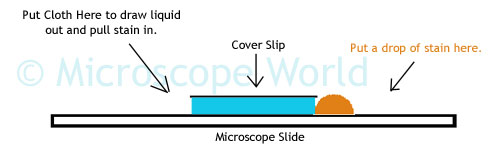
The image above shows how to draw a stain into a prepared slide. With the cover slip in place on top of the specimen, place a drop of stain on the edge of the cover slip. On the opposide side of the cover slip place a paper towel or cloth to draw the liquid out from the cover slip. As the liquid is drawn out, the stain will be pulled in under the cover slip.


