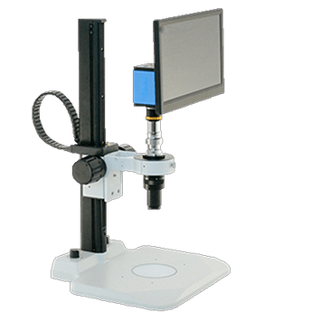
Ground US Continental Orders Over $200. Canada call for ship cost.
Phase Contrast Microscopes

Phase Contrast Microscopes

phase contrast microscopes Phase contrast microscopes constitute a vital component of modern biological research. The surfaces of glass plates within the body of phase microscopes are carefully scored to create rings. These rings induce a phase shift in the light passing through the medium being observed. Phase shifts create destructive interference patterns in the light that reaches the observer's eye. This method causes the observed image to appear out of normal phase by one half of one wavelength. Structures then become visible due to their contrast against a dark field behind them. Phase contrast microscopes allow researchers to observe differences between structures that have a similar level of transparency. By eliminating the need for time-consuming staining, phase microscopes allow research to be conducted more quickly and efficiently. 

For details regarding any of our featured microscopes, contact us. You can also request a quote today.

Phase Contrast Microscopes: A Breakthrough in Microscopy

Microscopy has revolutionized our understanding of the world we live in. From the smallest microbes to the intricate details of a butterfly wing, microscopes have opened up a world of discovery that is both fascinating and endlessly fascinating. One of the most important advancements in microscopy was the development of phase contrast microscopy.

What is a Phase Contrast Microscope?

A phase contrast microscope is a type of light microscope that enhances the contrast of transparent and semi-transparent specimens by using the principle of phase contrast. The phase contrast technique was developed by Dutch physicist Frits Zernike in 1932, for which he was awarded the Nobel Prize in Physics in 1953.

In traditional bright-field microscopy, specimens are viewed by light that passes through them and into the microscope objective. However, if a specimen is transparent or semi-transparent, such as living cells or tissues, they can appear nearly invisible under bright-field illumination. Phase contrast microscopy solves this problem by using a special optical system that converts the slight differences in phase of light waves passing through a transparent or semi-transparent object into differences in brightness or contrast.

How Does Phase Contrast Microscopy Work?

Phase contrast microscopy works by using a special phase contrast condenser that converts the light passing through the specimen into two beams, one that is in phase with the reference beam and one that is 180 degrees out of phase. These two beams are then recombined to form an image with enhanced contrast.

The phase contrast condenser consists of an annular diaphragm that is placed below the microscope stage and a phase plate that is placed in the objective lens. The annular diaphragm creates a hollow cone of light that illuminates the specimen obliquely, causing phase shifts in the light waves passing through the specimen. The phase plate then converts these phase shifts into intensity changes, creating a contrast image of the specimen.

Benefits and Applications of Phase Contrast Microscopy

The benefits of phase contrast microscopy are numerous. It allows for the observation of live, unstained specimens without the need for fixation or staining, which can alter the specimen's natural state. It also provides high-resolution images with excellent contrast and depth of field, making it ideal for studying the structures and functions of cells, tissues, and microorganisms.

Phase contrast microscopy is commonly used in biology and medicine for a wide range of applications, including the study of cell morphology, growth, and division, as well as the observation of microorganisms and viruses. It is also used in materials science and engineering to study the properties of transparent and semi-transparent materials, such as polymers and ceramics.

Phase contrast microscopy is a breakthrough in the field of microscopy that has greatly expanded our understanding of the microscopic world. Its ability to provide high-resolution, high-contrast images of transparent and semi-transparent specimens has made it an essential tool in biology, medicine, and materials science. With continued advancements in microscopy technology, we can look forward to even greater insights into the fascinating world of the microscopic.

Same Day Response

REQUEST A QUOTE

Submit Request

No Eyepieces Required

Visual Inspection Systems

Shop Now
Visual Inspection Systems
Loading...