
Monochrome microscopy cameras are sometimes overlooked, or often misunderstood as to what their benefit is for microscopy solutions. Monochrome microscopy cameras have a few benefits over color microscopy cameras:
- Produce sharper images with better resolution.
- Output smaller file sizes.
- More sensitive to light.
The main difference between a monochrome and a color image sensor is the lack of a color filter array (CFA). The removal of this optical band pass filter allows for a greater amount of photons to reach the photosensitive surface of the sensor, rendering it more sensitive to light which equates to a higher quantum efficiency. In addition, color cameras usually have IR cut filters to prevent infrared light from generating color aberrations. Without these filters, the red, green, and blue pixels would also react to various wavelengths in the NIR band resulting in the generation of strange and inaccurate colors. Since each pixel of a monochrome sensor can detect a broader spectrum of light due to the absence of the CFA and the IR cut filter, the overall performance of the camera improves significantly in low light conditions. Additional improvements are observed at higher wavelengths, specifically beyond 650nm, where Near Infrared (NIR) begins.
Microscopy cameras that are equipped with color filter arrays must interpolate the color data filtered out by the optical filter using a complex demosaicing algorithm. Because the data is interpolated and not measured, a margin of error is introduced. By removing the CFA, the added error is eliminated. The result is a sharper image with a monochrome camera and a higher effective resolution, since each pixel of the monochrome sensor contains a measured value that has not been influenced by its neighbor's value.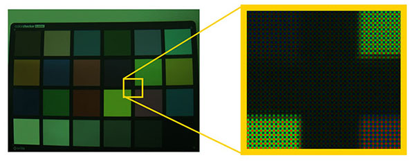
The image above is an enlargement of a raw RGB image of a color chart. The color channels are displayed as seen by the image sensor with Bayer pattern, without demosaicing.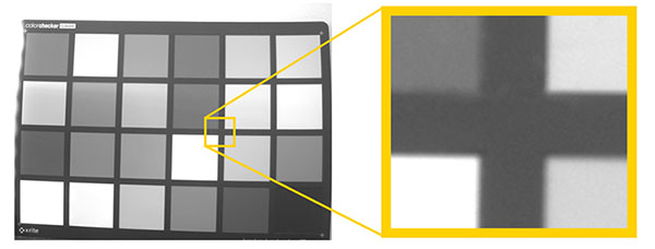
The image above is an enlargement of a raw monochrome image capturing the same color chart as above. Gray levels are consistent for each area and no Bayer pattern, no demosaicing required.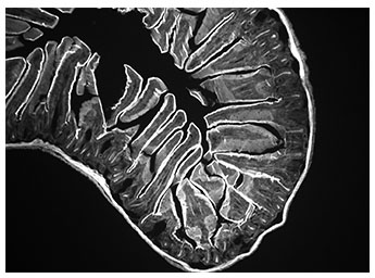 The removal of the CFA is responsible for the reduction of the monochrome image file size. Since monochrome image data does not contain specific color channel information, the image size is reduced threefold. This size reduction would also allow an increase in bit depth while maintaining storage savings, camera permitting.
In life sciences where microscopy specimens are stained with fluorescence emitting wavelengths above 650nm a monochrome microscopy camera is helpful.
This image is a mouse intestine sample that was captured under the microscope using the Lumenera Infinity 3-1M monochrome microscopy camera.


