Your shopping cart is currently empty.
IMA/USP Counting Chamber Microscope Reticle
The IMA/USP counting reticle can be purchased here.
IMA/USP Microscope Reticle that meets USP 788 Government Standards.
The IMA/UPS counting reticle is used for manual particle counting, most often in the pharmaceutical industry. Government guidelines require a manual microscope particle counting system must include a circular diameter reticle (installed in the microscope eyepiece) that is designed specifically for this purpose and calibrated using a certified stage micrometer.
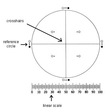
The particle counting reticle is divided into four quadrants by crosshairs - each showing transparent and black reference circles 10um and 25um in diameter at 100x magnification. The reticle also must contain a linear scale graduated in 10um increments (see below image).
 When manually counting particles the user will place the filter under the microscope with the counting reticle in the eyepiece. By carefully comparing any particles seen under the microscope to the reference circles, the user can record the number of particles of particular sizes (generally more than 10um and 25um in diameter). The user then marks this information in a workbook and will later create reports based on this data.
The reticle shown at left meets USP 788 standards for particle inspection. It has four symmetric quadrants, with transparent and black reference circles and a linear scale that is graduated in 10um increments.
You can order a IMA/USP counting reticle that meets USP 788 standards here.
Before placing an order for the IMA/USP reticle, you will need to know the diamter eyepiece reticle accepted by your microscope (if you are unsure please call or email us and we can help you determine this), along with the magnification factor (MF) of your microscope. The MF is unique for each microscope and 10x objective combination.
How to calculate your microscope's magnification factor (MF value):
- Set up the microscope with all the attachments and lenses you will use during the IMA. the IMA requires a compound microscope with two 10x eyepieces, a 10x objective lens, and episcopic (vertical) illuminator. IMPORTANT NOTE: In order to correctly determine the MF value, the vertical illuminator must be installed along with the other attachments affecting the microscope tube length/magnification (anything that is placed between the microscope objective and the eyepiece).
- Insert a 10mm / 100 division scale reticle into one eyepiece.
- Adjust the microscope eyepiece interpupillary distance.
- Adjust the magnification to 100x by moving the 10x objective into place.
- Turn on the microscope illuminator and focus the eyepiece so that the reticle lines are clear and sharp. Rotate the eyepiece diopter ring to focus, not the microscope focusing knob.
- Place a certified stage micrometer of 1mm / 100 divisions on the microscope stage and focus the microscope on the graduated lines.
- Adjust the microscope stage X and Y controls to align the stage micrometer parallel with the eyepiece reticle scale at the zero position.
- Record the largest number of whole eyepiece reticle scale divisions per corresponding whole stage micrometer divisions. To make the measurement as accurate as possible, a large part of each scale must be used. The ratio of eyepiece reticle scale divisions / stage micrometer divisions is the magnification factor (MF).
Example:
- If there are 75 reticle scale divisions per 80 stage micrometer divisions then MF = 0.938 or 75/80.
- If there are 95 reticle scale divisions per 92 stage micrometer divisions then MF = 1.032 or 95/92.
You can order a IMA/USP counting reticle that meets USP 788 standards here.