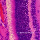
Retina
The retina is a light-sensitive layer of tissue that lines the inner surface of the eye. Learn more about the retina here.
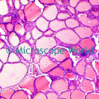
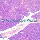
Human Thyroid Gland This cross section of human thyroid gland shows cuboidal and colloid stroma. The thyroid gland is one of the largest endocrine glands in the body. The thyroid is found in the neck, below the adam's apple. The thyroid gland controls how quickly the body uses energy, makes proteins, and controls how sensitive the body should be to other hormones. View an image of the entire thyroid gland here.
Human Liver Tissue Stained cross section of the human liver with white fibrous tissue. The liver is a vital organ with a wide range of functions including detoxification, protein synthesis, and production of biochemicals necessary for digestion. The liver is required for survival and it is not currently possible to live without a functioning liver. Learn more here.
Gall Bladder The gall bladder is a small organ where bile is stored before it is released into the small intestine. Humans can live without a gall bladder. Learn more here.
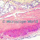
Human Esophagus
This cross section through the upper region of the esophagus shows the striated muscle. The esophagus is in the back of the throat and is an organ consisting of a muscular tube through which food passes to the stomach. View a diagram of the human esophagus here.
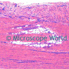
Human Tendon
This longitudinal section of a tendon shows strands of cells in dense fiber bundles. A tendon is a thick band of fibrous connective tissue that connects muscle to bone. A human tendon is capable of withstanding tension. The tendon is made of collagen. Tendons and muscles work together and can only exert a pulling force. Learn more here.
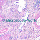
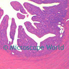
Columnar Epith
These are pseudo-stratified sections from the intestines. Epithelium is a tissue composed of cells that line the cavities and surfaces throughout the body. Learn more here.
Spleen The spleen is an organ in the upper part of the abdomen and acts as a filter for blood and in immune system health. View human spleen image here.
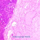
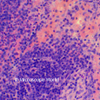
Human Pancreas
This cross section of the pancreas shows acini with secretory and centyrolinar cells and islands of Lengerhans. The pancreas is a gland organ in the digestive and endocrine system of vertebrates that produces important hormones including insulin, glucagon and somatostatin. It also secretes pancreatic juice containing digestive enzymes that pass to the small intestine. These enzymes help breakdown carbohydrates, protein and fat. Learn more here.