
When it comes to magnifying the microscopic world, a high-power or compound microscope stands as the ultimate tool, surpassing the capabilities of its stereo or low-power counterparts. Its prowess lies in revealing the intricate structures of smaller specimens, such as cell components, which remain hidden at lower levels of magnification. Within the framework of a compound microscope, a fascinating interplay of structural and optical components takes place. In the ensuing exploration, we will delve into the fundamental components that are essential knowledge for any enthusiast of microscopy. Illustrated and elucidated below are the pivotal microscope elements you'll want to acquaint yourself with for a deeper understanding of this remarkable instrument.
Compound Microscope Parts Labeled Diagram
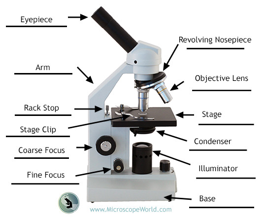
Compound Microscope Structural Components
1. Eyepiece (Ocular): The eyepiece, or ocular lens, is the lens closest to your eye when you look into the microscope. It further magnifies the image produced by the objective lens, allowing for a detailed examination of the specimen.
2. Arm: The arm is the curved part that connects the base of the microscope to the head. It provides support and stability to the microscope, and it is also where you grip the microscope when carrying it.
3. Rack Stop: The rack stop is a mechanism that limits how far up the stage (where you place the slide) can go. It helps prevent the objective lens from touching the slide and potentially damaging it.
4. Stage Clip: Stage clips are small clips located on the stage, designed to hold the specimen slide in place during examination. They ensure that the slide remains steady, allowing for accurate observation.
5. Coarse Focus: The coarse focus knob is used to make large adjustments to the focus of the microscope. It moves the stage up and down to bring the specimen into rough focus.
6. Fine Focus: The fine focus knob allows for precise adjustments to the focus of the microscope. It is used after coarse focusing to bring the image into sharp and clear detail.
7. Revolving Nosepiece (Turret): The revolving nosepiece holds the objective lenses and allows you to easily switch between them. It can be rotated to select different levels of magnification.
8. Objective Lens: Objective lenses are located on the revolving nosepiece and are responsible for the initial magnification of the specimen. Different objective lenses provide varying levels of magnification, typically ranging from low to high power.
9. Stage: The stage is the flat platform where you place the specimen slide. It often includes slide clips or a mechanical stage to secure the slide in place and move it smoothly for examination.
10. Condenser: The condenser is positioned beneath the stage and is responsible for focusing and directing light onto the specimen. It helps improve the clarity and brightness of the image.
11. Illuminator (Light Source): The illuminator is the light source of the microscope. It may be built-in or separate, providing the necessary illumination to illuminate the specimen for observation.
12. Base: The base is the lowermost part of the microscope, providing stability and support. It is the foundation upon which the rest of the microscope is built.


