Slide Mount Instructions
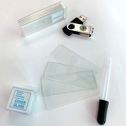
Before you begin the slide preparation process, ensure you have gathered all necessary materials, including slides, cover slips, droppers or pipets, and any chemicals or stains you intend to utilize.
There are two primary types of slides you'll be working with:
1. The standard flat glass slide.
2. The depression or well slide, which features a small well or indentation at its center designed to hold a drop of water or liquid substance. These are typically pricier and are commonly used without a cover slip.
Standard slides can be made of either plastic or glass, measuring 1 x 3 inches (25 x 75 mm) in size and 1 to 1.2 mm in thickness.
For wet slides, a cover slip or cover glass is essential. This is an ultra-thin, square piece of glass (or plastic) that is carefully positioned over the sample drop. Without the cover, surface tension would cause the droplet to form a dome shape. The cover counters this tension, ensuring the sample lies flat for precise examination with minimal focusing. Moreover, the cover acts as a safeguard, preventing the objective lens from interfering with the sample drop.
Microscope Slide Mounts
Microscope mounts refer to the methods used to secure microscope slides or specimens for observation. There are primarily four types:
1. Slide Mounts: These are the most common type and involve placing a specimen on a flat glass slide, often using a mounting medium (like a liquid or gel) to hold it in place. A cover slip is then added to protect the specimen and provide a flat surface for viewing.
2. Cavity or Well Mounts: These slides have a small well or depression in the center, designed to hold a drop of liquid, such as water or a specialized mounting fluid. They're used when working with specimens that require immersion in a liquid.
3. Temporary Mounts: These mounts are not meant for long-term storage or observation. They involve simple and quick methods like placing a specimen on a slide with a drop of water. They're often used for observing live organisms or time-sensitive samples.
4. Permanent Mounts: These are designed for long-term storage and observation. They involve more complex procedures, including the use of specialized mounting media that harden over time, encapsulating the specimen in a transparent medium. This type of mount is often used for educational slides and scientific research.
Each type of mount has its specific applications and is chosen based on the nature of the specimen and the purpose of the observation.
Four Common Ways to Mount a Microscope Slide
There are several common methods to mount a microscope slide, depending on the type of specimen and the purpose of observation. Here are four common ways to mount a microscope slide:
Dry Mounting:
Procedure: In this method, a specimen is placed directly onto a clean, dry microscope slide. It is often used for specimens that do not require additional moisture or immersion in a liquid.
Applications: Dry mounting is suitable for observing solid specimens like plant parts, hair, or minerals.
Wet Mounting:
Procedure: A small drop of liquid (usually water or a specialized mounting fluid) is placed on the slide, and the specimen is then added to this drop. A cover slip is gently placed over the specimen to prevent drying out and distortion.
Applications: Wet mounts are used for observing live organisms, cells, or specimens that require immersion in a liquid.
Squash Mounting:
Procedure: This technique involves placing a specimen on a slide and then gently pressing it down with a cover slip or another slide to flatten it. This is commonly used for soft tissues or delicate plant parts to create a thin, transparent layer for observation.
Applications: Squash mounts are used for examining tissues, especially in plant biology.
Smear Mounting:
Procedure: A small amount of the specimen (usually a liquid or semi-liquid substance) is spread thinly and evenly across a slide, often using the edge of another slide. It is then allowed to air-dry or may be fixed with a chemical agent.
Applications: Smear mounts are commonly used for observing microorganisms like bacteria or for preparing blood smears for microscopy.
These methods cater to different types of specimens and their specific requirements for observation. The choice of mounting technique depends on factors such as the nature of the specimen, the purpose of observation, and the desired level of detail and clarity.
Microscope Stains
Microscope stains, also known as biological stains or dyes, are substances used in microscopy to enhance the contrast and visibility of biological specimens. They work by selectively binding to specific cellular components, such as proteins, nucleic acids, or lipids, which allows for easier visualization and differentiation of cellular structures. Here are some common types of microscope stains:
1. Hematoxylin and Eosin (H&E):
Type: Combination stain.
Usage: One of the most widely used stains in histology, Hematoxylin stains cell nuclei blue-purple, while Eosin stains cytoplasm and some extracellular structures pink.
2. Methylene Blue:
Type: Basic dye.
Usage: Stains cell nuclei blue. It is commonly used for general cytology and microbiology studies.
3. Giemsa:
Type: Combination stain.
Usage: Used for staining blood cells, it differentiates nuclear and cytoplasmic components in a range of colors, allowing for the identification of various cell types.
4. Gram Stain:
Type: Combination stain.
Usage: Distinguishes between two major categories of bacteria: Gram-positive (which retain the crystal violet stain) and Gram-negative (which take up the safranin counterstain).
5. Wright's Stain:
Type: Combination stain.
Usage: Primarily used for blood smears, this stain allows for differentiation of blood cell types and identification of abnormalities.
6. Lugol's Iodine:
Type: Iodine-based stain.
Usage: Used in conjunction with Gram's iodine, it can be used to enhance the contrast of certain cellular structures.
7. Oil Red O:
Type: Lipophilic dye.
Usage: Binds specifically to lipids, making it useful for staining structures like fat droplets within cells.
8. Periodic Acid-Schiff (PAS):
Type: Chemical reaction-based stain.
Usage: Reacts with carbohydrates, highlighting structures like glycogen, mucins, and other glycoproteins.
9. Toluidine Blue:
Type: Basic dye.
Usage: Stains acidic components in cells and tissues, including mast cell granules and nucleic acids.
10. Safranin:
Type: Basic dye.
Usage: Used as a counterstain in Gram staining to color Gram-negative bacteria.
These are just a few examples, and there are many other specialized stains used for specific purposes in microscopy, such as special stains for connective tissues, nerve fibers, or specific cell structures. The choice of stain depends on the type of specimen and the information needed from the microscopic examination.
Microscope Staining Steps
1. Begin by preparing a wet mount slide, positioning your specimen on a clean microscope slide.
2. Using an eye dropper or pipette, carefully gather a droplet of the chosen stain.
3. Place this drop of stain on the outer edge of your cover slip, ensuring it contacts the slide's edge.
4. To facilitate the even distribution of the stain, position a piece of napkin or paper towel along the opposite side of the cover slip, pressing it firmly against the edge.
5. If necessary, apply an additional stain drop to guarantee complete coverage of your specimen.
6. Your slide is now fully prepared for observation under the microscope.
