

Microscopes for Reproductive Medicine
Nov 28th 2017
There are three types of reproductive medicine that use microscopes for fertility treatments in both humans and animals and each uses some different microscopy techniques. In all three reproduction techniques the eggs (ova) are removed from a female and fertilized outside of the female's body (in vitro) with sperm. Fertilized eggs (zygotes) are then implanted into the same or another female's uterus to establish pregnancy. The three reproductive medicine techniques are: In Vitro Fertilization (IVF), Intracytoplasmic Sperm Injection (ICSI) and Intracytoplasmic Morphologically-Selected Sperm Injection (IMSI).
In Vitro Fertilization (IVF)
The process of In vitro fertilization (IVF) starts with eggs being harvested and incubated. Next unselected sperm cells are added and both are left together (in vitro) for several days. During this time, healthy, mobile sperm cells actively fertilize the eggs. Finally, embryos are transferred into the uterus.
Intracytoplasmic Sperm Injection (ICSI)
The process of Intracytoplasmic sperm injection (ICSI) starts similar to IVF with eggs being harvested and incubated. Next the oocyte is stabilized by a holding pipette while a glass micropipette is used to collect a single sperm. This unselected sperm cell is immobilized by cutting its tail with the point of the micropipette. The oocyte is pierced through the membrane (oolemma) and the sperm is directed to the inner part of the oocyte (cytoplasm). The sperm is released into the oocyte. During this process the cellular structures such as the zona pellucida and polar body of the egg cell must be clearly visible.
Intracytoplasmic Morphologically-Selected Sperm Injection (IMSI)
Similar to both IVF and ICSI, the first step of IMSI involves harvesting and incubating the eggs. Next a morphological selection of a healthy egg is made using an inverted microscope. The oocyte is stabilized by a holding pipette. Semen is analyzed using an upright biological microscope. Morphological selection of a healthy sperm cell is made by using an nverted microscope with high magnification, DIC contrast and oil immersion objective lenses. A glass micropipette is used to immobilize the selected sperm by cutting its tail. The oocyte is pierced through the membrane (oolemma) and the sperm is directed to the inner part of the oocyte (cytoplasm). The sperm is then released into the oocyte. During this process, similar to ICSI, the cellular structures such as the zona pellucida and polar body of the egg cell must be clearly visible. The shape and vacuole count of the sperm cells must be assessed.
An upright microscope is primarily used for semen analysis and sperm preparation. Male infertility is diagnosed by detailed semen analysis that looks specifically at the total volume, number of motile sperm per milliliter (ml) and sperm morphology. Pre-selection of sperm takes place in glass dishes and requires a microscope with DIC or polarization.
Harvested cells are kept in culture dishes maintained at body temperature. Examination and selection of cells requires microscopes with heating stages. Initial sorting of cells is performed with a stereo microscope. Because egg cells sit at the bottom of the culture dish, in order to assess the cell's morphology, the objective lens must be close to the cells. This is why an inverted microscope is used for this stage. These microscopes also provide the room required for micromanipulation. For ICSI and IMSI the microscope system needs to be stable and vibration free. Special microscopy contrast techniques allow assessment of the morphology of unstained egg cells. In particular, ICSI and IMSI require high resolution and high contrast. A few of the microscopy techniques are listed below in detail.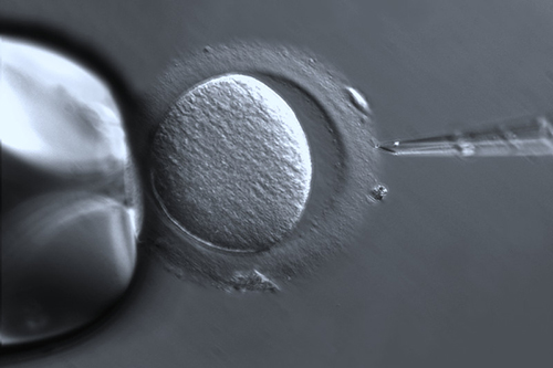 This is an image of an oocyte captured under the ZEISS Axio Vert.A1 inverted microscope using PlasDIC.
Differential Interference Contrast (DIC)
DIC provides high resolution images so that the finest structures in cells can be visualized. DIC uses polarized light to detect differences in the refractory index in specimens. Because plastic does not preserve the polarization of light, DIC will not work with a plastic dish.
PlasDIC
ICSI is often performed in plastic petri dishes, which as mentioned above, will not work with DIC. PlasDIC is a ZEISS technique that overcomes this limitation and delivers crisp relief contrast and an impressive three dimensional image.
Hoffmann Modulation Contrast (HMC)
HMC is the classic contrasting technique for unstained, low-contrast specimens. HMC delivers a crisp relief contrast and reveals even finest structures in cell nucleus, the nucleus shape and nucleoli.
Polarization Contrast
The sperm selection for IMSI is often performed in glass dishes where polarized light needs to be used.
Below are some recommended microscopes often used in reproductive medicine.
Daily lab applications including brightfield, darkfield, phase contrast or fluorescence:
Basic handling and sorting of egg cells and viewing injected egg cells:
Observe and manipulate specimens with a larger working distance for handling and sorting of eggs. Additionally, quick isolation of eggs before fertilization and assessing growing embryos.
Inverted microscope for micromanipulation and contrasting techniques including DIC:
To learn more about which microscope is best for your reproductive medicine application contact Microscope World.


