

Mushrooms under the Microscope
Nov 26th 2017
Mushrooms, also known as fungi or toadstools, are the spore-bearing fruiting body of a fungus. The mushroom typically grows above ground or on top of its food source (on a log, or out the side of a mossy tree). A mushroom has a stem and a cap along with gills. The gills of the mushroom are often examined under a microscope to identify the mushroom classification.
The terms mushroom and toadstool date back many centuries. Between 1400 and 1600 AD the terms mushrom, mushrum, muscheron, mousheroms, mussheron and musserouns were used. The French word mousseron is a reference to moss and may be at the root of the word mushroom.
The most important microscopic feature for identification of mushrooms is the spores. A mushroom's spores color, shape, size, attachment, and reaction to chemical tests often leads to identification.
The images below of mushroom spores were captured using a digital educational microscope.
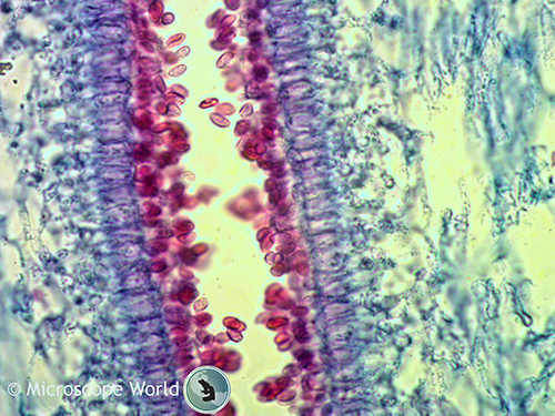
Edible mushrooms contain 20 percent or more daily value of B vitamins including riboflavin, niacin and selenium. This image of mushroom spores was captured at 400x magnification.
The term mushroom typically refers to the edible sporophores, while the term toadstool is sometimes reserved for inedible or toxic sporophores.
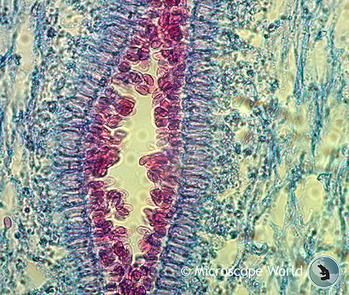
This image of a mushroom was captured at 100x magnification under a digital microscope.
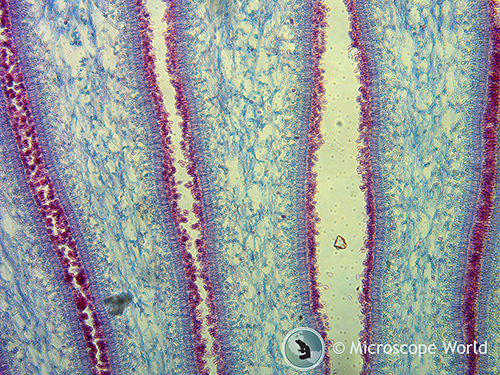
This image of mushroom spores was captured at 400x under a digital microscope.
Learn more about the history of mushrooms here. To learn more about microscopes and digital microscopy Contact Microscope World.


