DK3000 Microscope Digital Camera Images

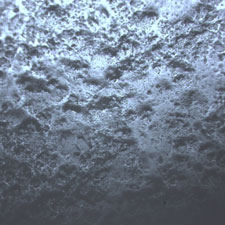
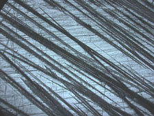
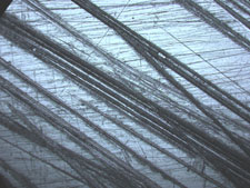
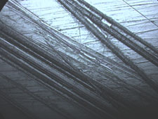
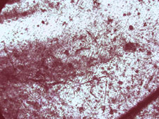
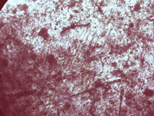
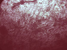
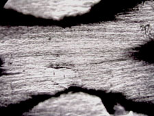
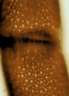
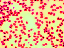
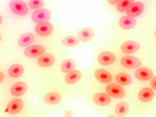
The images found below were all captured with the DK3000 3.1 mega pixel microscope digital camera that we carry. All images shown below were compressed to load faster on your computer, but are otherwise untouched photos.

Images below taken with the DK3000 3.1 mega pixel digital camera.

Arteries Image
  Image at left is of a human artery and was captured with the DK3000 3.1 mega pixel microscope digital camera. Click here to view the non-compressed version of the image.
Image shown at right is human tissue that was captured with the DK3000 3.1 mega pixel microscope digital camera. Click here to view the non-compressed version of the image.  
Tissue Image

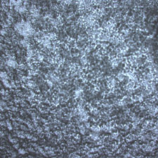
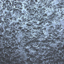
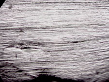
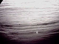
Images taken using the DK3000 3.1 mega pixel digital camera on the ML7100 Metallurgical Microscope

microscope image, aluminum 100x
microscope image, aluminum 200x
microscope image, aluminum 400x
Anodized aluminum, 100x
Anodized aluminum, 200x
Anodized aluminum, 400x
     
microscope image, steel, 100x
microscope image, steel, 200x
microscope image, steel, 400x
Scratched stainless steel, 100x
Scratched stainless steel, 200x
Scratched stainless steel, 400x
     
Microscope image, penny 100x
Microscope image, penny 200x
Microscope image, penny 400x
Penny, 100x
Penny, 200x
Penny, 400x
     
microscope image - plastic, 100x
microscope image - plastic, 200x
microscope image - plastic, 400x
Plastic strap, 100x
Plastic strap, 200x
Plastic strap, 400x

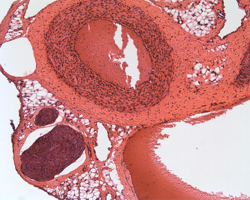
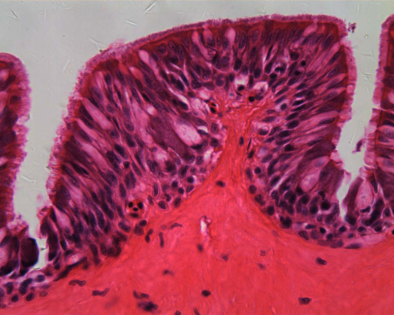
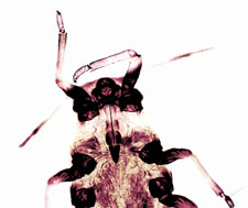
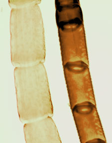
Images taken using the DK3000 3.1 mega pixel digital camera on the student biological microscope model 163

Microscope image of aphid
 

The image at the left is an aphid.

This image was captured at 100x magnification.

     
Microscope image, insect antenna 100x  

Image at the left are two different insect antenna. This image was captured at 100x magnification.


Image at right is the exact same antenna as that in the image on the left, but the image was captured at 400x magnification.

 
Microscope image, insect antenna, 400x
         
Microscope image of frog's blood, 400x  

Image at the left is frog's blood, notice the small dark dots in the center - these are the nuclei. This image was captured at 400x magnification.


Image at the right is the same microscope slide with frog's blood, but was captured at 1000x magnification. Now the nuclei of the blood cells are clearly visible.

  Microscope image, frog's blood, 1000x
Loading...