ProgRes® MF Series Camera Applications

The ProgRes® MF series cameras have a wide variety of applications. Below you will find images that were captured with some of the MF cameras with different microscopes.

Life Science
Fluorescence in Microbiology and Genetics:

metaphase FISH image

Sample:


Camera:

Source:

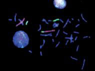
Metaphase; fluorescence in situ hybridization (FISH) is a cytogenetic technique that can be used to detdect and localize the presence of absence of specific DNA sequences on chromosomes. FISH is often used to find specific features in DNA. These features might be used for genetic counseling, medicine or species identification. Learn more about FISH here.

ProgRes® MFcool

Institute of Genetics; Clinical Center, Chemnitz, Germany

bovine pulmonary artery image


Sample:

Camera:

Source:

Bovine pulmonary artery endothelial cells; cytoskeleton. Cytoskeleton is the cellurlar skeleton contained within the cytoplasm. Learn more about cytoskeletons here.

ProgRes® MFcool

University of Chemnitz, Germany

bovine cytoskeleton

Sample:


Camera:

Source:

Bovine pulmonary artery endothelial cells; cytoskeleton. The endothelium is the thin layer of cells that line the interior surface of blood vessels. Learn more about endothelial cells here.

ProgRes® MFcool

University of Chemnitz, Germany

multi color fluorescence image

Sample:

Camera:

Source:

Multi color fluorescence

ProgRes® MF

ProgRes® application laboratory; Jena, Germany

Material Science
Quality Assurance:

carbon image

Sample:

Camera:

Source:

Carbon inclusion in steel

ProgRes® MF

Customer image

weld seam image

Sample:

Camera:

Source:

Weld seam

ProgRes® MFcool

Customer image

Metallography:

grain boundary image

Sample:

Camera:

Source:

Grain boundary

ProgRes® MFcool

ProgRes® application laboratory; Jena, Germany

Loading...