|
The ProgRes® cameras have a wide variety of applications. Below you will find images that were captured with either the C3 or C5 cameras with different microscopes including: stereo microscopes, biological microscopes and metallurgical microscopes.
|
| Life Sciences |
| Pathology |
|
|
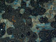
Sample:
Camera:
Source:
|
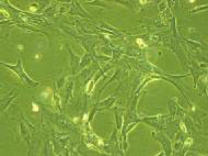
Human tumor cells. Learn how tumor cells form here.
ProgRes® C5
Institute of Laser Technologies in Medicine and Measurement; University Ulm, Germany
|
|
|
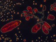
Sample:
Camera:
Source:
|
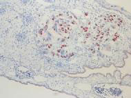
Glioblastoma cells. These are a type of primary central nervous system tumor, most commonly found in the brain, but can also be found in the spinal cord or optic nerves. Learn more here.
ProgRes® C5
German Cancer Research Center, Heidelberg
|
 |
Sample:
Camera:
Source:
|
Appendix
ProgRes® C5
ProgRes® application laboratory; Jena, Germany
|
| Biology |
 |
Sample:
Camera:
Source:
|
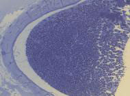
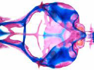
Rat section of sciatic nerve, click here to view the human sciatic nerve
ProgRes® C3
Electron Microscopic Center of the Medical Faculty; University Rostock, Germany
|
 |
Sample:
Camera:
Source:
|
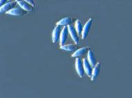
Gyromitras gigas 04-005 spores; 40er Varel-Contrast. Gyromitras gigas is a fungi, learn more about it here.
ProgRes® C3
Dr. Geert Schmidt-Stohn; Bienenbüttel, Germany
|
 |
Sample:
Camera:
Source:
|
Gyromitras gigas 04-005 spores; 40er Varel-Contrast. Gyromitras gigas is a fungi, learn more about it here.
ProgRes® C3
Dr. Geert Schmidt-Stohn; Bienenbüttel, Germany
|
 |
Sample:
Camera:
Source:
|
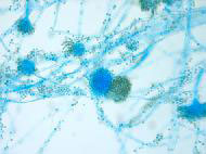
Aspergillus, learn more about aspergillus here.
ProgRes® C5
ProgRes® application laboratory; Jena, Germany
|
| Genetics |
 |
Sample:
Camera:
Source:
|
TIR fluorescence of adhered T47D-EYFP-Mem; breast cancer cells. Learn more about breast cancer here.
ProgRes® C3
Institute of Applied Research; University Aalen, Germany
|
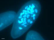 |
Sample:
Camera:
Source:
|
Live cells of the ciliate paramecium with DNA-selective dye Hoechst 33342 showing one stage of the macronuclear fragmentation through a sexuell process called autogamy.
ProgRes® C3
Institute of Biology; University Konstanz, Germany
|
| Material Science |
| Quality Insurance |
 |
Sample:
Camera:
Source:
|
Detail of a SQUID (superconducting quantum interference device)
ProgRes® C3
Max-Planck-Institute of Radio Astronomy; Bonn, Germany
|
 |
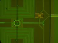
Sample:
Camera:
Source:
|
Circuit board viewed with a dual arm pipe illuminator under a stereo microscope
ProgRes® C3
ProgRes® application laboratory; Jena, Germany
|
 |
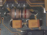
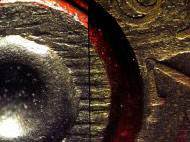
Sample:
Camera:
Source:
|
Hardness test effect. Interested in learning more? Click here for more information on material hardness and a variety of hardness tests.
ProgRes® C5
Customer photo
|
| Minerology, Geology, Material Research |
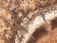 |
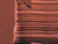
Sample:
Camera:
Source:
|
Concrete 30µm thin section, captured with transmitted light and bevelled polars
ProgRes® C3
Mike Eden; Geomaterials Research Services, Ltd.; UK
|
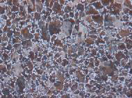
 |
Sample:
Camera:
Source:
|
Concrete 30µm thin section, captured with transmitted light and crossed polars
ProgRes® C3
Mike Eden; Geomaterials Research Services, Ltd.; UK
|
 |
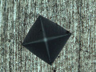
Sample:
Camera:
Source:
|
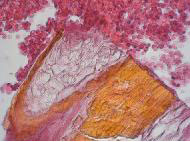
Cauixi, captured with brightfield
ProgRes® C3
Otto-Schott-Institute of Glass Chemistry; University Jena, Germany
|
| Metallography |
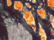
 |
Sample:
Camera:
Source:
|
C60 steel, captured with brightfield incident light
ProgRes® C3
ProgRes® application laboratory; Jena, Germany
|
 |
Sample:
Camera:
Source:
|
Gray cast iron with sphaerolithe, captured with brightfield incident light
ProgRes® C5
ProgRes® application laboratory; Jena, Germany
|
 |
Sample:
Camera:
Source:
|
Copper with copperoxidul
ProgRes® C5
ProgRes® application laboratory; Jena, Germany
|
| Forensics |
| Securing of Evidence |
 |
Sample:
Camera:
Source:
|
Impression of synthetics with same relief of abrasives
ProgRes® C5
Bavarian State Office of Criminal Investigation; Munich, Germany
|
 |
Sample:
Camera:
Source:
|
Breech face marks
ProgRes® C5
Bavarian State Office of Criminal Investigation; Munich, Germany
|
|
|