Prepared Slides: Musculoskeletal Histology Microscope Slides

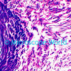
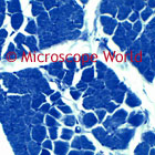
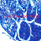
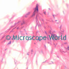
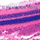
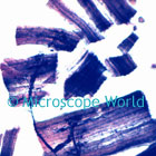
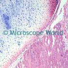
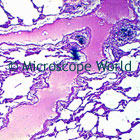
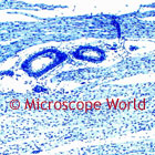
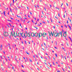
The images below were captured with the DMBA210 digital biological microscope. These prepared slides are included in the Histology Musculoskeletal Prepared Slide Kit.


Umbilical Cord image

Human Umbilical Cord
The umbilical cord connects a developing embryo or fetus to the placenta. In humans it normally contains two arteries and one vein. Learn more here.

Striated Muscle Microscope Image Striated Muscle
This cross section of striated muscle shows muscle fibers and bundles.
Spinal Cord microscope image Human Spinal Cord
This section of spinal cord shows gray and white matter. The spinal cord is a long, tubular bundle of nervous tissue and support cells that extends from the brain.
Smooth Muscle Microscope Image Smooth Muscle
These teased, isolate muscle fibers have been carefully stained to show pindle-shaped cells with single elongated nuclei.
Optic Nerve microscope image Optic Nerve
These cross section and longitudinal section nerve bundles show nerve fibers and connective tissue coverings.
motor nerve microscope image

Motor Nerve Ending
These motor nerve endings were teased and carefully selected to show both the branching nerve fibers and the end plates.

larynx microscope image Larynx
A longitudinal section showing cartilage and other topo-graphically related structures.
lung microscope image Human Lung
Cross section of lung showing thick cornified layer. The lung is an essential respiration organ in air-breathing animals. Learn more here.
cardiac muscle microscope image Cardiac Muscle
Section stained to demonstrate intercalated discs and approximate central location of nuclei in fibers.
aorta microscope image Aorta
This is a large artery section. The aorta is the largest artery in the body. It originates at the heart and extends down to the abdomen, where it branches off into two smaller arteries.
Loading...