Your shopping cart is currently empty.
Bacteriology Microscope Prepared Slides
All bacteriology microscope prepared slide images below were captured using the Richter Optica U2D digital biological microscope. Looking for a prepared slide kit? Check out our General Slide Kit.
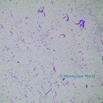 |
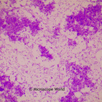
Bacillus Subtilis, Hay, Soil This bacteria is known as hay bacillus or grass bacillus. It is rod shaped and is found in the human intestines. The prepared microscope slide image of Bacillus Subtilis at left was captured at 400x magnification. Learn more here. |
|
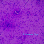 |
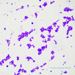
E. Coli E. Coli under the microscope at 400x. E. Coli (Escherichia Coli) is a gram-negative, rod-shaped bacterium. Most E. Coli strains are harmless, but some serotypes can cause food poisoning in their hosts. The harmless strains are part of the normal flora of the gut. Learn more about E. Coli here. |
|
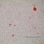 |
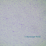
Helicobacter Pylori The prepared microscope slide image of Helicobacter Pylori at left was captured at 400x magnification. Helicobacter Pylori is a gram-negative bacterium linked to the development of duodenal ulcers and stomach cancer. However, over 80% of the individuals infected with Helicobacter Pylori are asymptomatic and it has been theorized that the bacterium may play an important role in natural stomach ecology. Learn more here. |
|
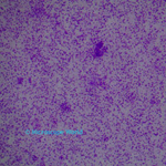 |
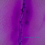
Lactobacillus (Yogurt Bacteria) Lactobacillus is a gram-positive rod-shaped bacteria, and is a part of the lactic-acid bacteria group. Most of the members of this bacteria group convert lactose to sugar and lactic acid. Lactobacillus bacteria that is found in the mouth (not from yogurt bacteria) can be responsible for tooth decay and dental cavities. The Lactobacillus microscope prepared slide image at left was captured at 400x magnification. |
|
 |
Rhizobium Meliloti (Nitrogen fixing in root on legume spear) Rhizobium Meliloti microscope prepared slide image at left was captured at 400x magnification. Also known as Sinorhizobium Meliloti, this gram-negative nitrogen-fixing bacterium forms a symbiotic relationship with legumes that results in a new plant organ termed a root nodule. |
|
 |
Sarcina Sarcina is a gram-positive bacterium that may be found on human skin or in the large intestine. The Sarcina prepared microscope slide at left was captured at 400x magnification. |
|
 |
Staphylococcus Staphylococcus microscope prepared slide image at left was captured at 400x magnification. This gram-positive bacteria is typically harmless and resides on the skin and in mucous membranes. Staph infections are caused by a strain of this bacteria. |
|
 |
Tuberculosis Tuberculosis is a disease that attacks the lungs and is caused by Mycobacterium Tuberculosis is a very weak gram-positive bacteria that can withstand disinfectants and survive in a dry environment for weeks. The microscopy image of Tuberculosis at left was captured at 400x magnification. Learn more about Tuberculosis here. |